Endometrial sampling: Endometrial carcinoma


Drs. Prolla and Diehl's INTERESTING CASE OF THE MONTH April 2002 case A

Papanicolaou staining

Papanicolaou staining

Papanicolaou staining

Papanicolaou staining

Low power 40x H&E staining

Medium power 100x H&E staining

High power 200x H&E staining

High power 500x H&E staining

High power 500x H&E staining

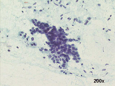
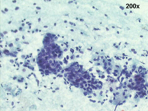
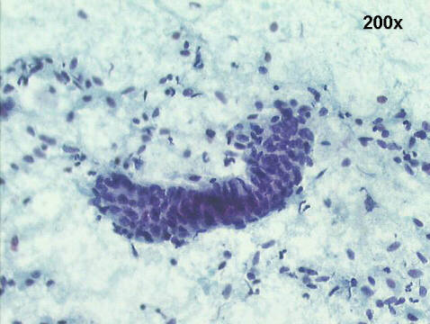
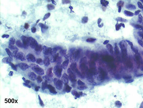
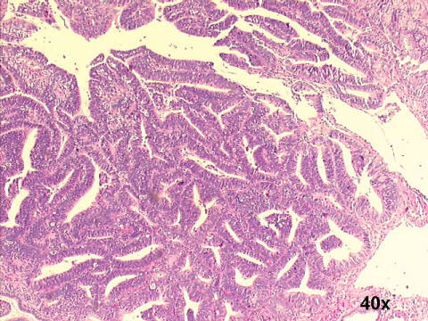
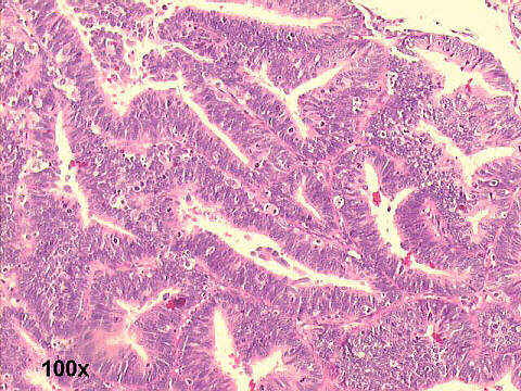
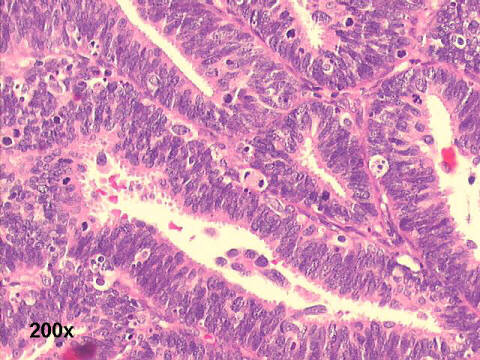
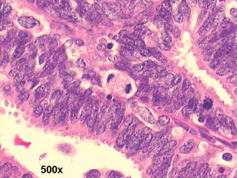
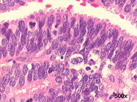
The cytology smears show high cellularity, large loosely cohesive sheets of cells, with palisade architecture, and nuclear crowding and hyperchromasia. The histology slides show well-differentiated endometrial adenocarcinoma, endometrioid differentiation. The recognizable glands are lined by stratified columnar malignant cells. Numerous mitotic figures, and apoptotic bodies, are also seen.


See also Case B


References
List of cases
Atlas
Hosted by www.Geocities.ws

1