Dr. Prolla INTERESTING CASE OF THE MONTH - March 2000
|
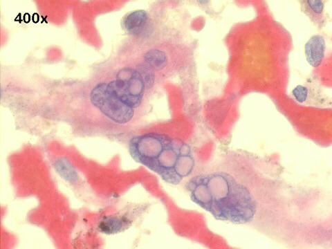
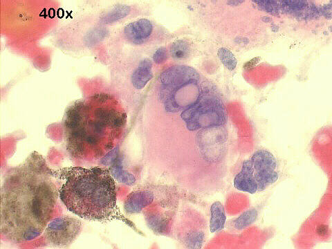
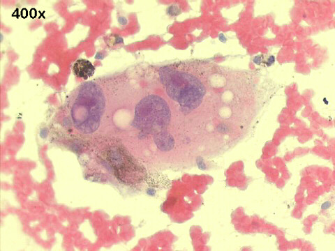
This is a typical case of metastatic malignant melanoma, with characteristic morphology: large and small single anaplastic cells (melanocytes) with intranuclear cytoplasmic invaginations, and finely dispersed cytoplasmic brown pigment ("dust like"), and histiocytes with aggregates of dark brown pigment (assumed to be melanin). The pigment can be seen also in the background. Spindle-shaped cells are also present. The nucleoli are usually prominent. Amelanotic melanomas are a diagnostic dilemma, and can be quite difficult to identify: poorly differentiated adenocarcinomas, large cell anaplastic Ki-1 lymphomas, and sarcomas being the main differential diagnosis. Rare cases of melanoma have spindle cells, and others have huge intracytoplasmic vacuoles (signet ring cell melanoma), easily mistaken for adenocarcinoma cells. Immunocytochemistry is almost always positive for S-100, and some cases are positive for vimentin. Most cases, about 80%, also stain with HMB 45, the so-called melanoma specific antigen (See Nasiell K, Tani E, Skoog L. FNA cytology and immunocytochemistry of metastatic melanoma. Cytopathology 1991; 2:137-147), with negative staining for epithelial markers. In difficult cases the melanin pigment must be confirmed by Masson-Fontana staining, and differentiated from hemosiderin ( with Perl's Prussian blue stain) and from lipofucsin (with PAS staining). As a last resource, electron microscopy will help demonstrating melanosomes.
Hospital de Clinicas de Porto Alegre, Pathology & Cytopathology
Laboratories
|

| References |
|