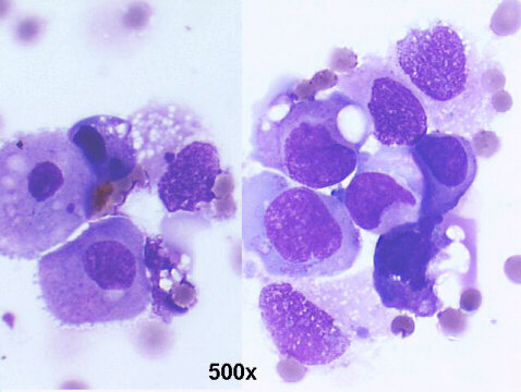
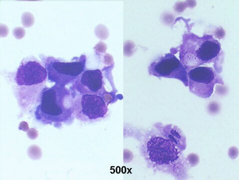
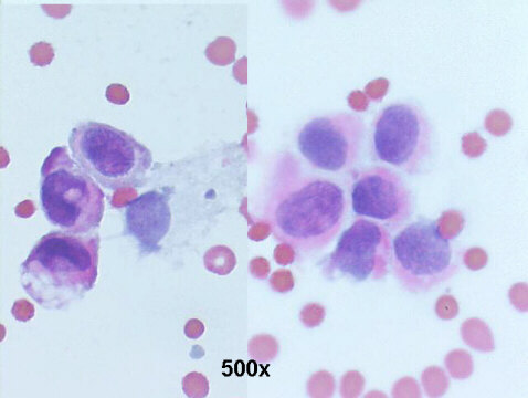
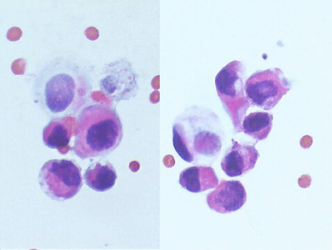
About 10% of the transitional cell carcinomas are carcinoma in situ (CIS) lesions. This is a flat type of tumor that spreads along the surface of the bladder and can progress to a muscle invasive tumor. CIS can be difficult to diagnose and can coexist with papillary tumors. CIS may have a characteristic red, velvety appearance when viewed endoscopically. At times, CIS is not visible and the diagnosis is made from cytologic examination of the urine or by obtaining random bladder biopsies. The cytologic diagnosis is made by the presence of numerous isolated urothelial malignant cells, on repeated urine samples, as seen in this case.
