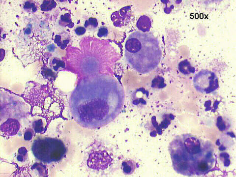
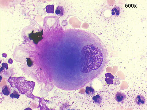
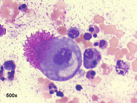
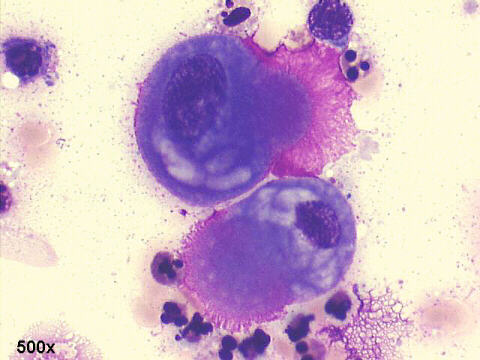
The presence of many microvilli in malignant cells is frequently documented by electron microscopy (EM), but their presence can be seen in some cases by optical microscopy as well. The use of Romanovsky stains can be very useful in this context ( I use to say they are the EM of the poor...). Spriggs and Boddington, in their classic book Cytology of effusions, Grune & Stratton, 1968, 2nd edition, were the first to call attention to this finding in papillary carcinomas of the ovary, with pictures remarkably similar to the ones in our case. More recently, this finding has been called "anemone cells" and "anemone tumors" in relation to the cells or the tumors bearing them, respectively