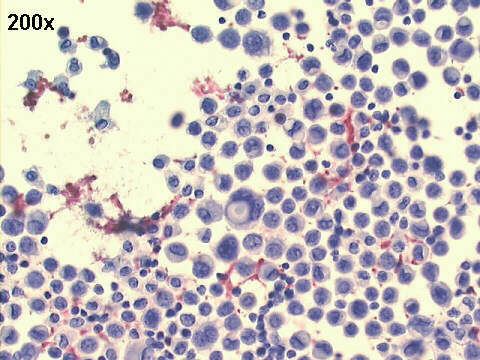
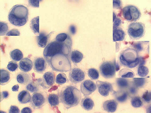
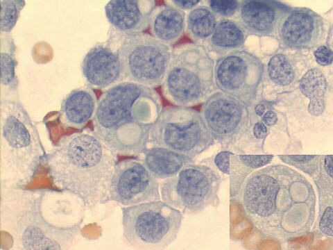
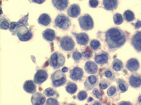
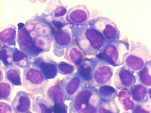
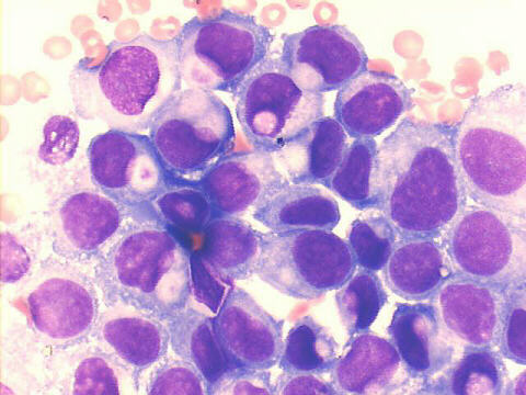
The smears show many cells which contain one or more intracytoplasmic lumens (ICLs). They are a very good criterion for malignancy, but they should be carefully distinguished from simple intracytoplasmic vacuoles. ICLs have well defined borders, and have dots or masses of inspissated mucin. Arthur I Spriggs & D W Jerrome (J Clin Pathol 28:929-936,1975) described such cells in effusions caused by breast carcinoma, and wrote: "The cells are characterized by the frequent presence of 'bull's-eye' vacuoles, in which a central spot is deeply stained by Giemsa, eosin or periodic acid-Schiff (PAS). In one of these it was shown by electron microscopy that the vacuoles are lined by microvilli". H. Battifora (Arch Pathol 99:614-617, 1975) confirmed these findings in histopathological material. They are very rarely found in benign breast lesions. ICLs have been described in effusions caused by many different types of adenocarcinomas, and one cannot infer the primary site from their presence.
Note: with this case we started to use a 20x oil immersion Planapo Olympus objective lens, with significantly better medium power images
|