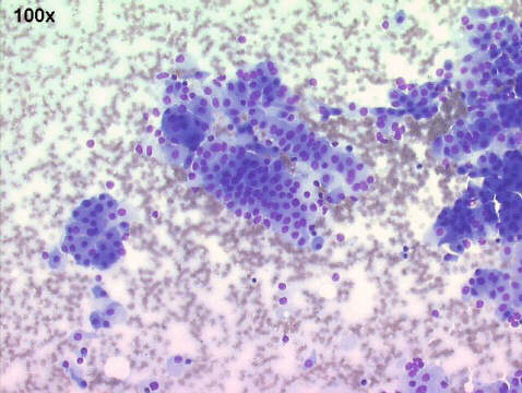
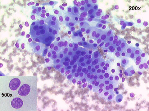
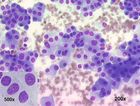
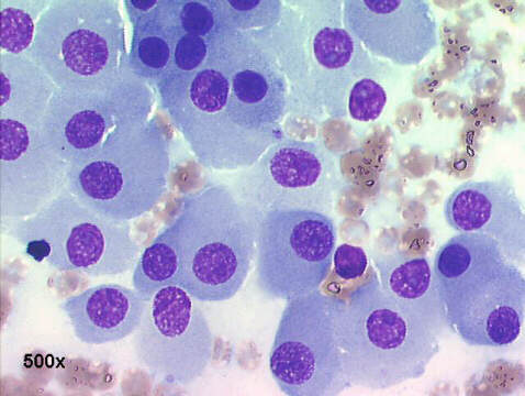
Lung oncocytoma: The cytology smears show the presence of many sheets of polygonal, oncocytic cells with abundant finely granular cytoplasm and centrally located round vesicular nuclei, most of them with a round prominent nucleoli. These cell are identical to the epithelial oncocytic cells seen in Warthin's tumor and oncocytomas of the salivary glands. The cytological diagnosis was oncocytic carcinoid versus lung oncocytoma.
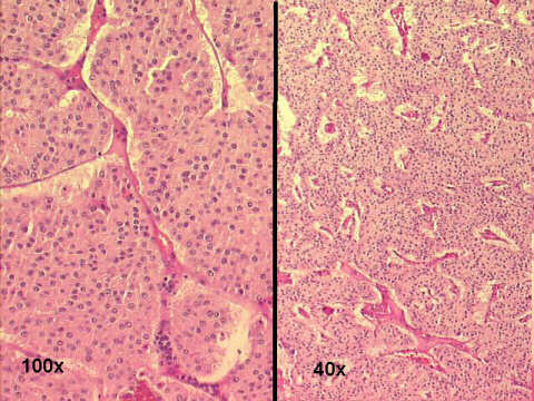
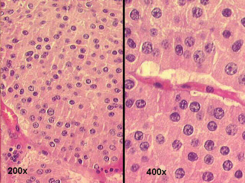
The histology of the tumor shows solid sheets or parallel columns of oncocytic cells, as described in cytology, with scarce fibrous septa. Mitoses and necrosis were absent. The H&E high power photos demonstrate the deeply eosinophilic and finely granular cytoplasm, and the round nuclei with prominent nucleoli. The sections were negative for neuroendocrine markers: chromogranin and synaptophysin, as well as for vimentin, and were positive for low and high molecular weight keratins. This pattern excluded an "oncocytic carcinoid" and the final histological diagnosis was the controverse "lung oncocytoma" or a mucous gland adenoma with oncocytic features (salivary gland type tumors of the bronchial tree).
See also Case A