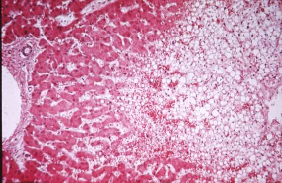
This is a classic nutmeg liver. It is due to chronic venous congestion secondary to right heart failure. The patient will also have congestion of kidneys, intestines etc and ankle edema. The darker areas in the gross picture are the enlarged vascular channels and the paler areas are the normal liver cells.
Slowing of blood flow through the sinusoids due to the back up from teh right heart, leads to imparied oxygen supply to the centrilobular cells and these may show fatty change. (see picture)
If prolonged some central cells may die and we may see the reticulin framework of the liver more clearly, also, there may be some deposition of fibrous tissue in this region. This finding is called Cardiac Cirrhosis. |