this image is interpreted by radiologists to provide a diagnosis.let us see the machines used in radiology
Xrays: they are the most basic equipment. xray is produced when fast moving electrons strike a tungsten target. the rays so produced "x-rays" penetrate the body and produce blackening in a silver bromide coated film. bones are visualised clearly because they do not allow much of the rays to reach the film. thus they are seen as white structures. xrays are very useful in diagnosing fractures and bone tumors
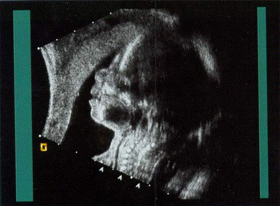
ultrasound scan: here sound waves of high frequency are transmitted into the body and the reflected sound is converted into electrical signals and displayed in a screen. since they use sound they are safely used in pregnancy to know the position of the fetus, to calculate the gestational age of fetus and to detect fetal defects.currently3D and 4D scanners are available which produce more realistic images
CT scans: here multiple cross sectional images are produced with the aid of computer hence called Computed axial tomography or CAT scan. it gives images of any part of the body including the brain where ultrasound has difficulty in reaching. they are primarily used to see hemorrhage in brain , to decide operability of tumors etc
MRI scan: here magnetic field 10000 times of earths field is applied and the signal changes are analysed by a computer. they are used for diagnosis of tumors, spinal cord injury etc and also for research