Docent Dr Dragan Sagic
Dedinje Cardiovascular Institute
M. Tepica 1 Str,
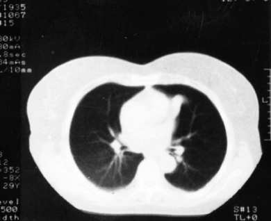
YU- 11000 Beograd,Yugoslavia
E- mail: [email protected]
remark: on-line version of this article is still incomplete
DISCUSSION
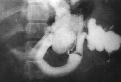
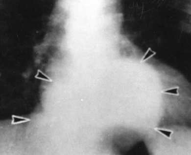
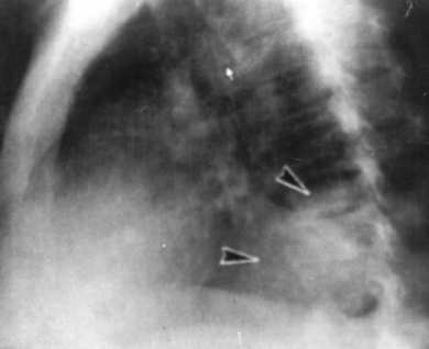
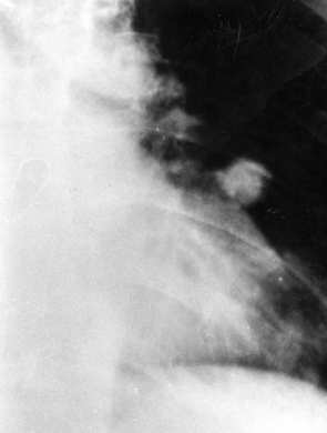
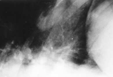
Mediastinal shadow enlargement and posterior mediastinal opacifications on the standard chest x - rays in patients with portal hypertension, are caused by dilatation of one of the large veins (azygos or hemiazygos), or by dilatation of smaller, mediastinal veins (paraesophageal varices) (1,6). The incidence of visualization of PV on the standard chest radiograms is 5-8% (1,4). Higher sensitivity (11%) was noted by applying Ishikawas "Bucky" technique (4). Relatively high incidence of visualization of PV was result of careful analysis of the radiograms and cocomparisons with the angio- and manometry records. Lee suggests use of CT of the thorax, since the visualization of the mediastinal structures is better (3). False positive interpretations are mostly caused by the erroneous judgement of the obliteration of the lower right paraspinal space in the presence of the pleural effusion, cardiomegaly, elevated right hemidiaphragm and elongated thoracic aorta (3,4). Widening of the mediastinal shadow may be present in different clinical conditions such as oesophageal diseases¸ enlargement of the paraesophageal lymph nodes¸ other posterior mediastinal tumorous masses etc.
Differential diagnosis of PV is sometimes very difficult in patients with lymphomas, which have enlarged paraesophageal lymph nodes and splenomegaly. In this cases, CT or MRI are very valuable (3). Interspace of the descending thoracic aorta may be obliterated (or may have nodulated aspect) in the presence of paraoesophageal lesions such as mediastinal lymphadenopathy, hiatal hernia, retroperitoneal tumours spread and other conditions (3).
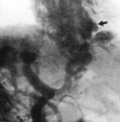
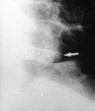
Dilated azygos and/or hemiazygos vein, as well as large PV, may change the contour and the density of the posterior mediastinum, being described as mediastinal pseudotumours (3, 8, 9). In this series of 140 patients, we found only one case of mediastinal pseudotumour, caused by the dilatation of the hemiazygos vein. Figure 4. Since PV was not detected, this case was not included in this study. In his group of patients Moult found one case of the dilated azygos vein. On the contrary Lee detected higher incidence of isolated dilatations of azygos (20%) and hemiazygos vein (4%) (1¸3).
Clinical importance of the visualization of the PV on the standard chest radiograms seems to be very high, since this may reveal patients with long-lasting portal hypertension, or those in the terminal stadium of the portal hypertension. On the contrary Guermazi did not find any correlation between the degree of the enlargement of the oesophageal varices and the number and the size of the paraoesophageal collaterals (9). However¸ this patients need more variceal injection sessions and longer hospitalization (3, 9).
In conclusion, standard chest radiography is another method of detecting paraoesophageal verices. Enlarged mediastinal shadow with opacification of the posterior mediastinum along with splenomegaly may indicate the presence of the paraoesophageal varices. To avoid erroneous diagnosis of the mediastinal pseudotumors in patients with portal hypertension, additional CT, ultrasonography and portography are be helpful.