THE ROLE OF ULTRASOUND SCANNING OF THE LOWER ABDOMEN IN THE DIFFERENTIAL DIAGNOSIS OF ACUTE APPENDICITIS
Uloga ultrazvucnog pregleda donjeg dela trbuha u diferencijalnoj dijagnozi akutnog apendicitisa
( accepted December 23rd 1999 )
Anastasios C. Souparis, John G. Makris , Maria Arvaniti , Aris Patsas, Wasileios T. Papaziogas , John Galatianos , Theodoros E. Pavlidis, Thomas W. Papaziogas
2nd Surgical Clinic of Medical Faculty of the Aristoteles University of Thessaloniki,Thessaloniki, "G.GENNIMATAS" Hospital , Thessaloniki, Greece.
Address correspondence to:
Dr Wasileios Papaziogas
Bl.Gabriilidi 29
546 55 Thessaloniki¸ Greece
FAX ( 30 31 ) 200 212
E - mail [email protected]
Ultrasonography in acute appendicitis Soupras AC et al.
ABSTRACT
The aim of this prospective study was to assess the value of the lower abdominal ultrasonography in the diagnosis of acute appendicitis. Sixty patients ( 50 females and 10 males; mean age: 36.7 years ) with presumed acute appendicitis enrolled this study during 22-month period ( January 1997 - October 1998 ). The ultrasound findings were unequivocally positive in 41/45 patients, false negative in 3/45 patients and false positive in 1/45 patients. We therefore postulate that the abdominal ultrasonography is very useful diagnostic tool in acute appendicitis, differentiating it from other diseases of the lower abdomen.
Key words: acute appendicitis, appendiceal diseases, diagnosis, ultrasound
SAZETAK
Cilj ovog prospektivnog ispitivanja je bio da se proceni dijagnosticka vrednost ultrazvucnog pregleda donjeg dela trbuha u slucaju akutnog apendicitisa. ezdeset pacijenata ( 50 mukarca¸ 10 zena; prosecna dob: 36.7 godina ) sa sumnjom na akutni apendicitis je ukljuceno u ovo ispitivanje koje je trajalo 22 meseca ( Januar 1997 - Oktobar 1998 ). Ultrazvucni nalaz je bio nedosmisleno pozitivan u 41/45 pacijenata¸ lazno negativan u 3/45 pacijenata¸ i lazno pozitivan u 1/45 pacijenata. Na osnovu navedenog zakljucili smo da je utrazvucni pregled abdomena znacajna dijagnosticka metoda u slucaju akutnog apendicitisa.
Kljucne reci: akutni apendicitis¸ bolesti apendiksa¸ dijagnoza¸ ultrazvuk.
INTRODUCTION
Acute appendicitis might have various clinical manifestations. It may simulate almost any acute abdominal illness and in turn, may be mimicked by a variety of conditions. The diagnosis of acute appendicitis is mostly based on clinical evaluation ( lower right abdominal pain, rebound tenderness, leukocytosis etc ) as there are no other accurate diagnostic procedures with high specific diagnostic yield. Furthermore, in certain groups of patients such as children, elderly people, or pregnant women, the differential diagnosis of acute appendicitis could be even more difficult. The introduction of the ultrasonography has created new perspectives. This method in experienced hands has a high accuracy and may be a very helpful tool in the differential diagnosis of lower abdominal pain, especially in young females.
The 2nd Surgical Clinic of Aristotelian University of Thessaloniki and the Department of Radiology of " G. Gennimatas " Hospital Thessaloniki present their experience with abdominal ultrasonography in the diagnosis and differential diagnosis of acute appendicitis.
PATIENTS AND METHODS
Sixty patients were were enrolled this study over a 22 month period from January 1997 to October 1998. There were 50 females and 10 males with a mean age of 36.7 years ( range: 15-78 years). All patients presented at the emergency department of our clinic with acute lower right abdominal pain and/or leukocytosis setting the clinical suspicion of acute appendicitis. In these patients a graded compression real-time sonography of the lower abdomen was performed.
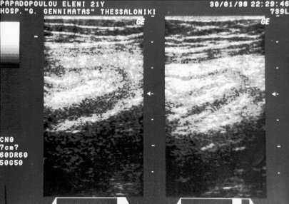
All sonograms were carried out by the same radiologist using a 3.5 MHz linear array transducer. The transducer directly visualised an inflamed appendix after all gas and fluid had been gradually swept out of the caecum and ascending colon by compression ( 1 ). When an appendix was not recognized immediately, oblique, transverse, and longitudinal scans of the right lower quadrant were made. A sonogram was interpreted as indicative of appendicitis when one - wall thickness of the visualized compressed appendix was greater 2mm or when appendiceal total outer wall-to-wall diameter was greater than 6 mm. Sonographic findings were interpreted as normal if after careful examination the appendix was not seen or, if seen, each single-wall thickness was less than 2mm and the outer wall-to-wall diameter was less than 6mm ( 2,3,4 ). Figure 1. The decision for laparotomy was made under careful consideration of clinical¸ laboratory¸ and sonographin findings.
The intraoperative and histopathologic findings of the specimens of the appendectomies were compared with the results of each sonogram.
RESULTS
45 out of the 60 patients underwent laparotomy ( 75% ). In all but two patients the diagnosis of acute appendicitis was proved intraoperatively ( 95.5 % ). In 2 patients appendix was not inflamed
Fourty out of 43 patients with intraoperative findings of acute appendicitis had a preoperative positive ultrasound ( 93% ). In the remaining 3 patients preoperative sonogram were negative ( false negative ).
Of the 2 cases with negative intraoperative findings for acute inflammation of the appendix, one had also a preoperative negative sonogram, and the other one had a positive finding for appendicitis. ( false positive ).
Fifteen patients ( 13 females and 2 males ), did not undergo laparotomy. These were usually patients with mild clinical signs, in whom the severity of the findings could not justify the indication for appendectomy. They were admitted in our department and received intravenous antibiotics, having a daily measurement of the leukocytes, until the clinical signs resolved. All patients showed a gradual improvement in a few days, so that no appendectomy was necessary to be performed. Thirteen of these patients had a negative sonogram, however two of them ( 13.3 % ) had a positive sonogram for acute appendicitis.
DISCUSSION
Acute appendicitis is the most common indication of emergency abdominal surgery. When the typical clinical signs are unequivocal, the decision to perform an operation is straightforward. However, many patients present with atypical right lower abdominal pain not characteristic for appendicitis. In these cases, the use of an imaging method, capable of showing changes of acute appendicular inflammation is of great importance. The graded-compression ultrasonography of the lower abdomen is a fast, sensitive and inexpensive diagnostic modality to confirm or rule out acute appendicitis. ( 4,5,6,7 ) Other, less specific and more expensive methods are CT, NMR or technetium 99 HMPAO scanning ( 8,9 ). They have great value in children, in whom sometimes the clinical symptoms are difficult to evaluate. Carrico et al reported that in his series sonographic findings resulted in revised clinical diagnosis of acute appendicitis in 52% of examined children ( 10,11 ).
The use of graded - compression ultrasonography is also valuable in differential diagnosis of acute lower right abdominal pain in pregnants, in whom the clinical diagnosis is oftenly difficult. Lim et al reported that the sonographic findings indicating acute appendicitis was confirmed in 93.5% of cases at operation. However, the use of compression sonography has to be performed carefully in pregnants in the third trimester. In this case Lim et al suggested left lateral decubitus position ( 12 )
Another advantage of the ultrasonograhy, is the fact that specialist such as a surgeon could not necessarily perform it. Amgwerd et al reported that the sensitivity and specificity of the ultrasonograms made by an experienced surgeon for the diagnosis of acute appendicitis were 97% and 98% respectively (13). Zielke et al reported that the overall accuracy and sensitivity of the clinical diagnosis of acute appendicitis were 84.9% and 51.3%, and those of ultrasounds performed by surgical residents 93.6% and 83.1%, respectively ( 14 ). Throughout evaluation of the results of clinical and ultrasound findings further improved the diagnostic performance ( accuracy 93.4%, sensitivity 84.1%, specificity 96.2%) and significantly reduced the rate of diagnostic errors to 3.4%. This is to suggest that the use of ultrasounds in differentiating acute appendicitis is not a diagnostic tool limited to experienced radiologists.
In conclusion we postulate, that in acute appendicitis abdominal ultrasonography performed by experienced clinicians have high diagnostic accuracy. This is of great help in differentiating patients with obscure abdominal symptomatology.
REFERENCES
1.Puviaert JBCM, Rutgers PH, Lalisang RI, et al. A prospective study of ultrasonography in the diagnosis of acute appendicitis. N Engl J Med 1987; 317: 666 -9.
2.Larson JM, Peirce JC, Ellinger DM, et al. The validity and utility of sonograph of appendicitis in the community setting. Am J Radiol 1989; 153: 687 -91.
3.Abu-Yousef MM, Bleicher JJ, Maher JW, et al. High-resolution US in the diagnosis of acute appendicitis Am J Radiol 1987; 149: 53 -8.
4.Jeffrey RB, Laing FC, Lewis FR. Acute appendicitis : High-resolution real-time US findings. Radiology 1988;167: 327 - 9.
5. Chang WM, Lee CH, Chou YH, et al. A clinical evaluation of ultrasonography in the diagnosis of acute appendicitis. Surgery 1989; 105: 154 - 9.
6.Simonovsky V. Sonographic detection of normal and abnormal appendix. Clin Radiol 1999; 54: 533 - 9.
7.Smajer B, Horalek F, Chvatalova N. Differential diasnosis of pain in the right lower abdominal quadrant. Rozhl Chir 1999; 78: 72 -5.
8.Loley CR, Latimer RG, Rimkus DS. Detection of acute appendicitis by technetium 99HMPAO scanning. Am Surg 1992; 58: 761 -5.
9.Incesu L, Coskun A, Selcuk MB, et al. Accute appendicitis : MR imaging and sonographic correlation. Am J Roentgenol 1997; 168: 669 - 74.
10.Lessin MS Chan M, Catallozi M, et al. Selective use of ultrasonography for acute appendicitis in children. Am J Surg 1999; 177: 193 -6.
11.Carrico CW, Fanton LZ, Taylor GA, et al. Impact of sonography on the diagnosis and treatment of acute lower abdominal pain in children and young adults. Am J Roentgenol 1999, 172 513 - 6.
12.Lim HK, Bae SH, Seo GS. Diagnosis of acute appendicitis in pregnant women: value of sonography. Am J Roentgenology 1992, 159: 539 - 42.
13.Amgwerd M, Rpthlin M, Candinas D, et al. Ultrasound diagnosis of appendicitis by surgeons: a matter of experience? A prospective study. Langebecks Arch Chir 1994; 379: 335 -40.
14.Zielke A, Hasse C, Sitter H, et al. Surgical ultrasound in suspected acute appendicitis. Surg Endosc 1997; 11: 362 -5.