http://www.collembola.org/images/gallery.htm
-
Last updated on
2009.09.15
by Frans Janssens
Preface
This is a collection of the most exclusive images on Collembola.
The images are kindly made available by its authors. Note that
the copyright of the images remains with the respective authors.
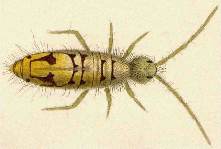
Lithographs
Reproductions of colourfull paintings of Collembola,
made by Hollick, A.T. in Lubbock, J. (1873),
courtesy of Gordon Ramel, 1995:
Entomobryidae: Entomobrya nivalis Entomobryidae: Willowsia buski More reproductions can be found at
Gordon Ramel's Collembola Pictures gallery .
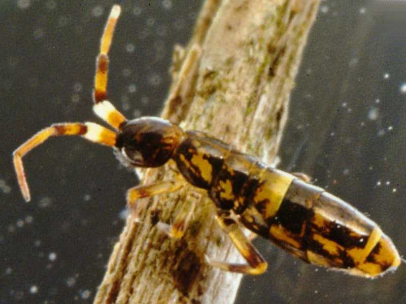
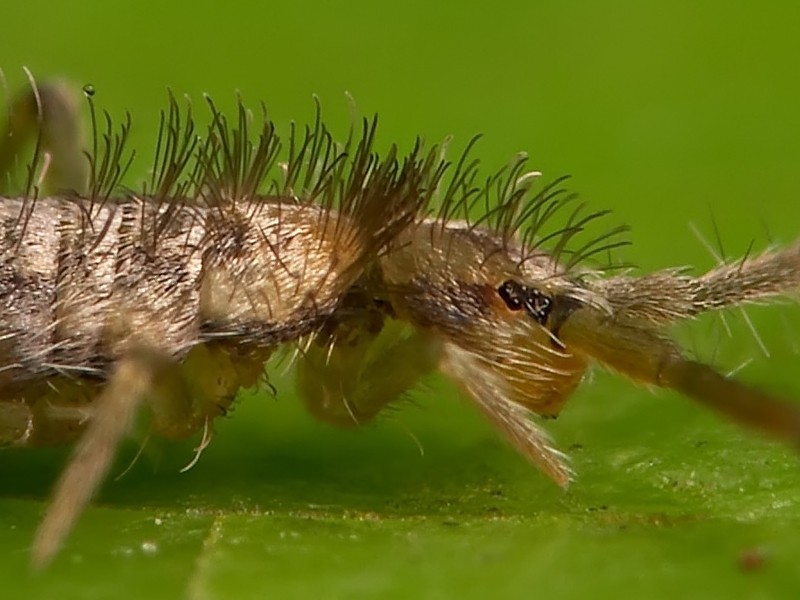
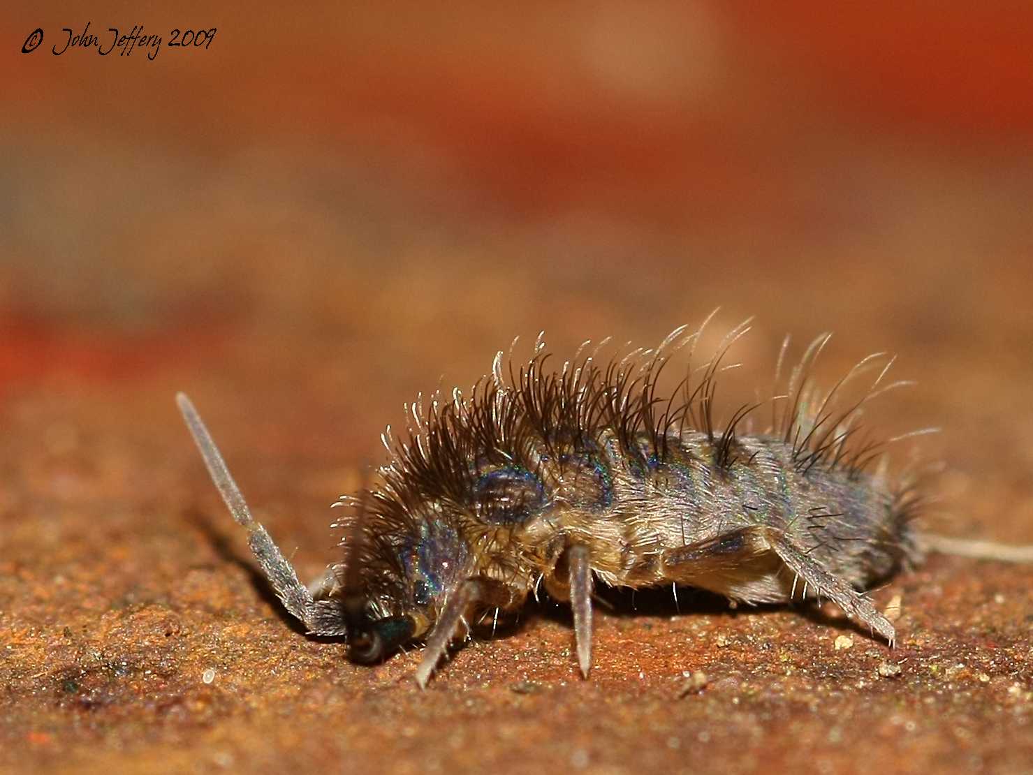
Macrophotographs
Remarkable photographs mostly of live specimens, courtesy of
Ab H. Baas,
Enrique Baquero,
Toby Barton,
Noel J. Cornwall,
Miroslav Deml,
Xavier Domene,
Mike Feldman,
Krister Hall,
Hans Henderickx,
Jarmo Holopainen,
Steve Hopkin,
John Jeffery,
Rafael Jordana,
Frithjof Kohl,
David R. Maddison,
Jonathan Schmidt,
Mark Stevens,
Brian Valentine
and
Bev Wigney
:
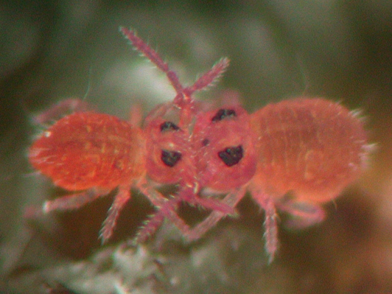
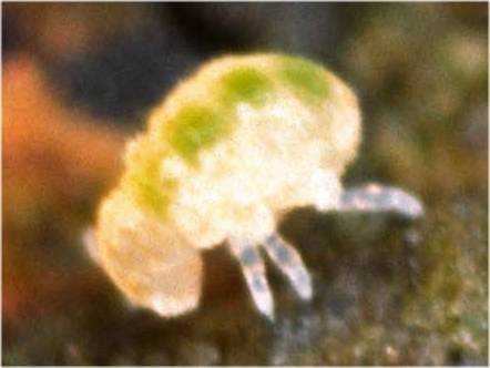
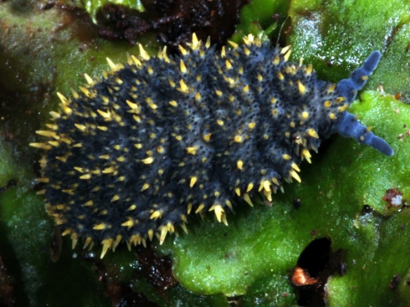
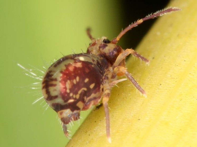
Hypogastruridae: Ceratophysella? Hypogastruridae: Ceratophysella armata Hypogastruridae: Hypogastrura nivicola Hypogastruridae: Hypogastrura viatica Neanuridae: Acanthanura sp. Neanuridae: Anurida granaria Neanuridae: Anurida granaria Neanuridae: Neanura muscorum Neanuridae: Bilobella aurantiaca Neanuridae: Holacanthella duospinosa giant springtail of 17 mm, habitus dorsal, © Stevens, 2007.
Onychiuridae: Tetrodontophora bielanensis Poduridae: Podura aquatica Isotomidae: Folsomia candida Isotomidae: Isotoma viridis Isotomidae: Isotomurus plumosus 2 Entomobryidae: Entomobrya intermedia Entomobryidae: Orchesella cincta Entomobryidae: Orchesella cincta Entomobryidae: Orchesella flavescens Entomobryidae: Orchesella flavescens Entomobryidae: Orchesella villosa Entomobryidae: Orchesella villosa Entomobryidae: Orchesella villosa Entomobryidae: Orchesella villosa Entomobryidae: Orchesella villosa Entomobryidae: Orchesella villosa Entomobryidae: Lepidocyrtus paradoxus Tomoceridae: Tomocerus vulgaris Tomoceridae: Pogonognathellus longicornis Tomoceridae: Pogonognathellus longicornis Neelidae: Megalothorax minimus Pogonognathellus longicornis , the giant of 6 mm, © Hopkin, 2005.
Arrhopalitidae: Arrhopalites hirtus Sminthuridae: Allacma fusca Sminthuridae: Vesicephalus europaeus Sminthurididae: Sphaeridia serrata Sminthurididae: Sminthurides aquaticus Dicyrtomidae: Dicyrtoma fusca Dicyrtomidae: Dicyrtoma fusca Dicyrtomidae: Dicyrtomina saundersi Dicyrtomidae: Dicyrtomina saundersi More fascinating photographs of live Collembola can be found at
Arthur Anker's photo gallery Springtails and Bristletails ,
Tristan Bantock's photo gallery Collembola from the UK, North of London ,
Toby Barton's photo gallery Springtails from the UK, Richmond Surrey ,
Toby Barton's other photo gallery Springtails from the UK, Richmond Surrey ,
Lynn Bergen's photo gallery Springtails from the USA, New York ,
Peter Birch's photo gallery Collembola-Springtails from the UK, Knutsford ,
Joe Botting's photo gallery Collembola from the UK, South of London ,
Peter Bryant's photo gallery Poduromorpha from the USA, California ,
Alistair Campbell's photo gallery Springtails from the UK, South of Birmingham ,
Arthur Chapman's photo gallery Springtails from Australia, Melbourne ,
Coder's photo gallery Springtails from the USA, Michigan ,
Noel Cornwall's photo gallery Springtails from the UK, Sussex ,
Rick Cowen's photo gallery Springtails from the USA ,
Rick Cowen's photo gallery Collembola from the USA, South Dakota ,
Alison Edwards's photo gallery Springtails from the UK, Wales, Port Talbot ,
Shane Farrell's photo gallery Springtails from the UK, Cheshire ,
Mike Feldman's photos and videoclips Springtails from the USA, Illinois, Champaign ,
Phil 'Goldenorfe's photo gallery Springtails from the UK, Thurstaston ,
Raphaël Haentjens' photos on Collembola from Belgium, Wallonie ,
Krister Hall's photo gallery Collembola from Sweden ,
Liz Henwood's photo gallery Springtails from the UK, East Sussex ,
Ronny Hermans's photo gallery Springtails from Belgium, Antwerp ,
Kathie Hodge's photo gallery Collembola from the USA, New York ,
Steve Hopkin's photo gallery Collembola from the UK, Reading ,
Gabor Keresztes's photo gallery Collembola from Central Europe: Hungary and Croatia ,
Gabor Keresztes's photo gallery Collembola from Japan ,
Brian Kilford's photo gallery Springtails from the UK, Wombourne ,
Michael Kilner's photo gallery Collembola from the UK, Bolton ,
Frithjof Kohl's photo collection Springtails from Germany ,
Pavel Krásenský's photo gallery Collembola from Czechia ,
Philippe Lebeaux's photo gallery Collemboles from France ,
J. LeMons' photo gallery Springtails from the USA ,
Stéphane Losacco's photo gallery Collemboles from France ,
Gary McDonald's photo gallery Collembola from the USA, California ,
Jonathan Michaelson's photo gallery Collembola from the UK, Berkshire ,
Maria Minor & Alastair Robertson's photo gallery Collembola from New Zealand ,
Cheryl Moorehead's photo gallery Springtails from the USA, Washington, Seattle ,
Tom Murray's photo gallery Springtails from the USA, Massachusetts ,
Alby Oakshott's photo gallery Springtails from the UK, Portsmouth ,
Tim Ranson's photo gallery Springtails from the UK, Channel Islands ,
Andrew Robertson's photo gallery Springtails from the UK, Scotland, Penicuik ,
Shadowshador's photo gallery Collembola from the UK, Wales, Park Hall ,
Gordon Spears' photographic forum on Collembla from the UK, Lancashire, Chorley ,
Gordon Spears' photo gallery on Collembla from the UK, Lancashire, Chorley ,
Mick Talbot's photo gallery Dicyrtomidae from the UK, Lincolnshire ,
Mick Talbot's photo gallery Entomobryinae from the UK, Lincolnshire ,
Mick Talbot's photo gallery Hypogastruridae from the UK, Lincolnshire ,
Mick Talbot's photo gallery Orchesellinae from the UK, Lincolnshire ,
Mick Talbot's photo gallery Tomoceridae from the UK, Lincolnshire ,
Scott Thompson's photo gallery Springtails from the UK, Somerset ,
Maarten Tonsbeek's photo gallery Springtails from the UK, Canterbury ,
José Ramón Pato Vicente's photo gallery Collembola from Spain ,
Brian Valentine's photo gallery Springtails from the UK, South Coast, Worthing ,
Lance van de Vyver's photo gallery Collembola from New Zealand, Levin ,
Michel Vuijlsteke's photo gallery Collembola from Belgium, Ghent ,
Phil Warner's photo gallery Collembola from the UK, St Albans , and
Bev Wigney's photo gallery Springtails from Canada, Ontario .
Bugguide's photo gallery Collembola from the USA and Canada ,
BioLib's photo gallery Collembola from Czechia ,
Monde des Insectes' photo gallery Collembola from France ,
The Great Smoky Mountains National Park photo gallery Collembola ,
Flickr's photo gallery Springtail ,
Flickr's photo gallery Springtails ,
Flickr's photo gallery Collembola ,
Flickr's photo gallery Springstaart ,
Rick Cowen's photographers showcase on Collembola/Springtails from the world .
Photomicrographs
Fascinating phasecontrast photomicrographs of morphological characteristics of Collembola,
courtesy of
Keith Brocklehurst:
Isotomidae: Tetracanthella brachyura , anal spines dorsal, © Brocklehurst, 1999.
Entomobryidae: Orchesella villosa , head lateral, © Brocklehurst, 2005.
Tomoceridae: Tomocerus minor , mucro lateral, © Brocklehurst, 1998.
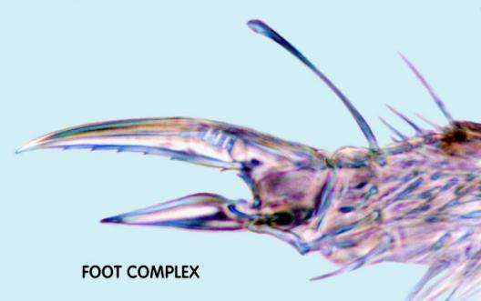
Tomoceridae: Tomocerus minor , footcomplex lateral, © Brocklehurst, 1998.
Sminthurididae: Sminthurides malmgreni , habitus lateral, © Brocklehurst, 1999.
More high quality photomicrographs of morphological features of the Collembola can be found at the
Springtail of the Month
pages of the PMS Springtail Study Group.
Interesting dark field photomicrographs of morphological characteristics of Onychiuridae,
courtesy of
Terry Lynch:
Onychiuridae: Onychiurus sp. , anal spines dorsal, © Lynch, 2001.
Onychiuridae: Onychiurus sp. , cuticular aspect dorsal, © Lynch, 2001.
Visit
Bioluminescent springtails light up Christchurch, New Zealand
by Terry Lynch (2001-)
for more detailed photomicrographs of morphological features of the
Onychiuridae and other Collembola.
High quality preparation microscope photomicrographs of the habitus of Collembola,
courtesy of
Arne Fjellberg:
Sminthurididae: Sminthurides aquaticus Dicyrtominae: Dicyrtoma fusca
Scanning Electron Micrographs
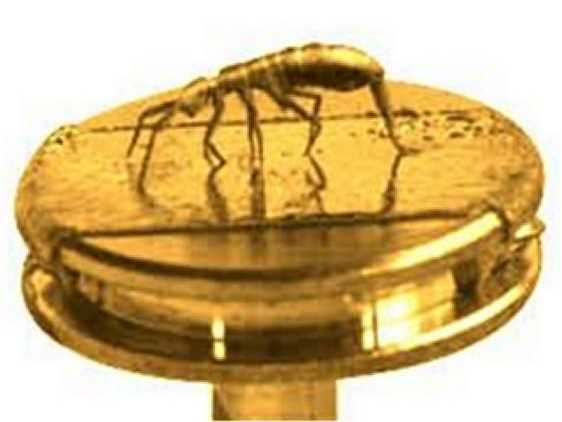
A gold sputtered specimen of Orchesella cincta , mounted on SEM stage by Hans Henderickx,
photographed by Stephane Vandermeeren and digitally postprocessed by Wilfried Hooftman:
Entomobryidae: Orchesella cincta
Morphological features of Isotomurus ,
courtesy of
T.M. Driscoll:
Isotomidae: Isotomurus Isotomidae: Isotomurus
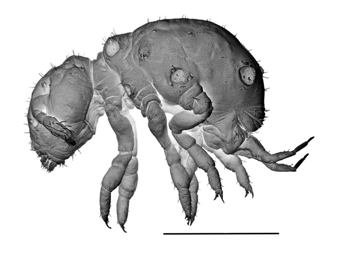
Stunning SEM's of family representatives,
courtesy of
David Walter and Raphael Jordana:
Entomobryidae: Heteromurus Isotomidae: Folsomides Neelidae: Megalothorax Bourletiellidae
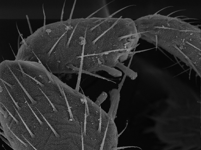
Amazing close-ups of the epicuticular ultrastructure,
courtesy of
Stéphan Borensztajn,
Helen Ghiradella and
Neil Plant:
Collembola Collembola Collembola Collembola Tomoceridae: Tomocerus Tomoceridae: Tomocerus flavescens?
Characteristic features of Actaletidae , Coenaletidae , and Sminthuridae ,
courtesy of
José Palacios-Vargas, and
Douglas Zeppelini:
Actaletidae: Spinactaletes sp. Actaletidae: Spinactaletes sp. Actaletidae: Spinactaletes sp. Coenaletidae: Coenaletes caribaeus Coenaletidae: Coenaletes caribaeus Sminthurididae: Sminthurides Sminthurididae: Sminthurides Sminthurididae: Sminthurides Sminthuridae Sminthuridae
A new species of Seira , with unique raptorial prolimb modifications,
courtesy of Douglas Zeppelini:
Entomobryidae: Seira
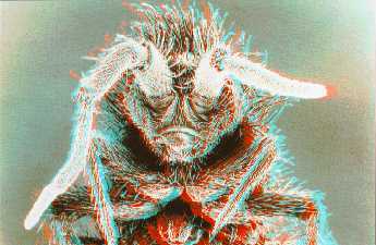
Three Dimensional Images
2D anaglyphs :
3D glasses 1 A dramatic face á face stereoscopic scanning electron micrograph,
courtesy of David Burder:
Entomobryidae: Orchesella
Cross-eye stereograms :
Some fascinating stereoscopic macrophotographs, courtesy of Brian Valentine
and Mike Feldman:
Hypogastruridae: Ceratophysella sp. Entomobryidae: Entomobrya intermedia Entomobryidae: Entomobrya intermedia Entomobryidae: Entomobrya intermedia Entomobryidae: Entomobrya intermedia Dicyrtomidae: Dicyrtomina ornata Dicyrtomidae: Dicyrtomina ornata Dicyrtomidae: Dicyrtomina ornata Dicyrtomidae: Dicyrtomina ornata Dicyrtomidae: Dicyrtomina ornata Dicyrtomidae: Dicyrtomina saundersi Dicyrtomidae: Dicyrtomina saundersi Dicyrtomidae: Dicyrtomina saundersi Dicyrtomidae: Dicyrtomina saundersi
Brian Valentine's Springtails Photo Gallery from the UK , and
Mike Feldman's photos and videoclips Springtails from the USA .
Progressive and regressive morph between Neanura and Dicyrtoma
Neanuridae: Americanura mexicana & Sphaeridia sp., foraging behaviour, clip of 1m10s, © Palacios-Vargas , 2005.
Neanuridae: Neanura muscorum Onychiuridae: Protaphorura armata Isotomidae: Isotoma viridis ?, nervously jumping, clip of 0m06s, © Holopainen, 2001.
Neelidae: Megalothorax? sp.Schmidt , 2005.
It is quite intriguing, because the "stop and head nod" behaviour is also
quite common in this species and may be a method of scanning for olfactory cues.
Arrhopalitidae: Arrhopalites hirtus Schmidt , 2005.
Bourletiellitidae: Deuterosminthurus bicinctus Sminthurididae: Sminthurides sp., mating behaviour, clip of 1m35s, © Palacios-Vargas , 2005.
Sminthurididae: Sminthurides sp., mating behaviour, clip of 1m24s, © Palacios-Vargas , 2005.
You also might want to visit
video clips
of pest and beneficial insects; or
Soil Animal Video clips .
You thought Collembola were not inspiring for artists, didn't you?
Sminthuridae: Sminthurus You might want to visit the unique
Collembola Art gallery
of artist Goetz Kluge.
Endnotes
1 How to obtain some 3D red-blue anaglyph glasses?
3D glasses can be ordered from companies such as
Deep Vision 3D
3D Glasses Direct
2 Tentative identification by Arne Fjellberg .