ULTRASOUND NOTES
HOW ARE ULTRASOUND WAVES PRODUCED?
Ultrasound waves are sound waves at frequencies above the upper limit of human hearing, which is 20,000 Hz.
Medical ultrasound is usually a lot higher in the 1 to 20 millions of Hertz range.
But, in order to make waves at this frequency you need a ‘loudspeaker’ that will vibrate at this frequency.
If you tried to get a normal speaker to vibrate a million times a second it would self-destruct!
So, what do you do?
You use a piezo-electric material.
A what?
A piezo-electric material.
These are crystals like quartz and lead zirconate titanate (PZT). These all have the special property that when they are squashed (or stretched) an electric voltage appears across them:
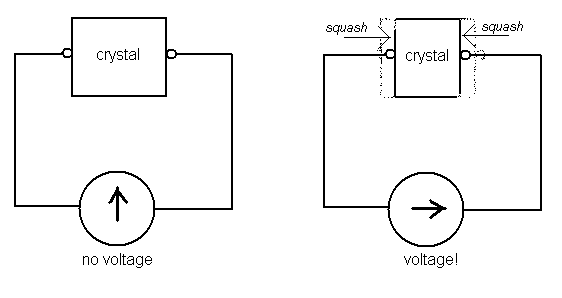
Basically the crystal is converting the mechanical energy of the squash into electrical energy.
These crystals are used in tiny key-ring torches where the energy used squashing the ‘on’ button is converted to electricity to power the bulb.
And in lighters where the squashing energy is converted to a high voltage that creates a spark that lights the gas.
The crystals can also be used as tiny microphones, because as sound waves ‘squash’ (or stretch) the crystal this creates a voltage that can be detected and processed by electronics.
And so, these crystals can also work the other way around, converting electric energy to mechanical energy.
If a voltage is put across a piezo-electric crystal it causes the crystal to squash (or stretch)
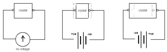
And if you put an A.C. voltage across the crystal then the crystal will squash and stretch in rhythm with the changing voltage – it will ‘vibrate’.
And if the A.C. voltage changes direction 1,000,000 times a second then the crystal will change shape 1,000,000 times a second, creating a sound wave with a frequency of 1,000,000 times a second – a million hertz!
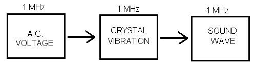
So, the frequency of the electric voltage controls the frequency of the sound waves produced – neat!
The actual device used in medical ultrasound is called an ‘ultrasound transducer’.
‘Transducer’ means a device that converts energy from one form to another.
Q)
Which forms of energy does an ultrasound transducer convert?
A) Electric energy to sound energy.
And remember that a transducer also converts sound energy
to electric energy…
Because when the echoes (sound waves) are received back by the transducer they are converted into electric signals that can then be processed by electronics.
So, an ultrasound transducer produces ultrasound waves AND it detects ultrasound waves (the echoes).
The way it works is that it sends out a pulse of ultrasound waves. Then it stops and listens for echoes. Then it sends out another pulse of waves. Then it stops to listen for echoes etc.
An medical ultrasound transducer
Here is a diagram of the transducer:
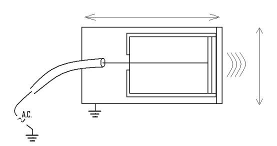
Here are the labels for the diagram:
q live electrode to apply voltage to crystal
q piezoelectric crystal that vibrates as voltage changes across it
q cable holding live wire
q earth connection to complete electric circuit
q ultrasound waves produced by transducer
q plastic window to protect crystal
q backing material to damp vibrations in crystal and live electrode
q sound insulator to stop sound hitting crystal from any other direction than through window
q 2 cm
q 5 cm
q alternating voltage
Copy the diagram and write in the labels in the appropriate place using your knowledge and intelligence!
THE MATHEMATICS OF ULTRASOUND TRANSDUCERS
Does it matter what the actual frequency of the ultrasound is?
Yes.
Because as you know:
- low frequency waves tend to spread out more, but get absorbed less so can travel further
- and high frequency waves tend to travel straighter, but get absorbed quicker so cannot travel as far.
And it is the same for ultrasound. If you want a wave to travel a long way into the body you need it to be a low frequency ultrasound wave, but if you want it to travel very straight so that it can give you better measurements of position you want it to be a high frequency ultrasound wave.
Penetration of ultrasound
Here is a table that gives typical penetration depths for different ultrasound frequencies:
|
Frequency (Hz) |
Penetration
depth (cm) |
Resolution (mm) |
Structures
investigated |
|
3-5 |
10-20 |
1.0 |
Deep: heart, uterus, liver |
|
4-10 |
5 |
0.2 |
Near surface: thyroid, carotid artery, breast |
|
10-15 |
1 |
0.1 |
Surface: eye |
|
50 |
Few mm |
0.05 |
Skin or surgical investigations (blood vessel walls, cartilage) |
Resolution of ultrasound
You can also see from the table that as the frequency of the waves increases the resolution (how small you can ‘see’/measure) gets better.
As with any wave there exists a relationship between the frequency of the wave, its wavelength (distance between waves) and the speed the waves travel at. The relationship is:
Speed of wave = frequency of wave
x wavelength of wave
Or
wavelength of wave = Speed
of wave / frequency of wave
The smallest that a wave can measure, the smallest distance that an ultrasound picture can ‘see’, is twice the wavelength of the ultrasound.
The speed of ultrasound in human tissue is about 1500 m/s.
So, for a 1 MHz wave the wavelength can be found by using the formula above:
Wavelength = 1500 / 1000000 = 0.0015 metres or 1.5 mm.
So, the smallest object a 1 MHz wave could resolve or ‘see’ would be a 2 x 1.5 mm = 3mm object.
Q)
What size objects can a 3 MHz ultrasound wave resolve?
A) Work it out!
Fundamental frequency of wave emitted by crystal
You can alter the frequency of the ultrasound waves by altering the frequency of the driving voltage but it is actually easier to use the transducer crystal like a ‘bell’ – you whack it with a high frequency pulse of electricity and this sets it ‘ringing’ at its own, fundamental frequency.
The preferred frequency that the crystal likes to vibrate at depends on the thickness of the crystal.
The relationship is expressed in the following formulae:
Preferred wavelength of emitted wave = 2 x thickness of
crystal
Or
thickness of crystal =
Preferred wavelength of emitted wave / 2
And
Preferred frequency of emitted
wave = 1900
/ thickness of crystal
Or
thickness of crystal = 1900 / Preferred
frequency of emitted wave
These are all the same formula, just re-arranged in different ways.
So, to produce a 1 MHz wave you want a crystal with a thickness of…
Required thickness of crystal = 1900/1000000 = 0.0019 m = 1.9 mm or about 2mm
Q)
What crystal thickness would you need to make a 10 MHz wave?
A) Work it out!
DOPPLER ULTRASOUND
The Doppler Effect is the principle behind police radar guns, behind blue-shift and red-shift for those of you who read SF and why sirens change pitch as they approach you and then move away from you.
QUICK EXPLANATION OF DOPPLER EFFECT
We know how sound waves echo off objects and so let you know that the object is there. Yes?
Well, what if the object is moving? What difference does it make?
Well, you still get an echo, but now the frequency of the echo is changed from the frequency of the original sound waves.
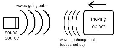
If the object is moving towards you then the echoed sound waves are ‘squashed up’ by the moving object and so are at a higher frequency than before.
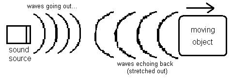
And if the object is moving away from you then the echoed sound waves are ‘stretched out’ by the moving object and so are at a lower frequency than before.
So, by studying how the frequency of the ultrasound changes you can actually measure how fast and in what direction the object is moving.
This is the basis of Medical Doppler Ultrasound which is ultrasound used to study moving objects inside the body, mainly the blood flowing through the veins and arteries.
The objects are the blood cells and just as in the diagrams above they echo back the ultrasound at different frequencies to give the doctor information about how they are moving.
Here is a diagram of how it works in practice:
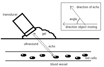
Obviously the original ultrasound and echo are not usually directly in line with the direction the object is moving, so the angle between the two directions affects the measurements.
The formula that relates all the factors involved is:
Change of frequency
= 2
x the original frequency x the speed of the object x cos of
the angle
the speed of the ultrasound waves
Q)
What would be the frequency change when ultrasound reflects off blood cells
travelling at 1 m/s, in an artery at 60o to the skin surface, using
waves of 5 MHz frequency that travel at a speed of 1500 m/s?
A) Work it out!
SET-UP OF A DOPPLER TRANSDUCER
The difference in the set-up of a Doppler transducer as compared to a normal transducer is in the electronic processing, because in the Doppler transducer you are not just interested in when there is an echo but you want to know the change of frequency of the echo.
Here is the diagram of the Doppler set-up:
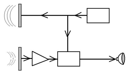
And here are the labels to accompany the diagram:
q transmitting crystal sending out high frequency wave
q receiving crystal detecting high frequency echo
q oscillator producing high frequency
q amplifier to boost echo to same loudness as transmitted signal
q electronics to subtract echo away from transmitted signal
q final Doppler signal played through loudspeaker
Copy the diagram and write in the labels in the
appropriate place using your knowledge and intelligence! Think about what might
be happening…