The ear is the sense organ that detects sounds. The vertebrate ear shows a common biology
from fish to humans, with variations in structure according to order and species. It not only
acts as a receiver for sound, but plays a major role in the sense of balance and body position.
The ear is part of the auditory system.
The word ear may be used correctly to describe the entire organ or just the visible portion. In
most animals, the visible ear is a flap of tissue that is also called the pinna. The pinna may be
all that shows of the ear, but it serves only the first of many steps in hearing and plays no role
in the sense of balance. In people, the pinna is often called the auricle. Vertebrates have a pair
of ears, placed symmetrically on opposite sides of the head. This arrangement aids in the
ability to localize sound sources.
In biology, an organ Latin: organum, instrument, tool is a group of tissues that perform a
specific function or group of functions. Usually there is a main tissue and sporadic tissues. The
main tissue is the one that is unique for the specific organ. For example, main tissue in the
heart is the myocardium, while sporadic are the nervous, blood, connective etc.
Introduction to ears and hearing
Audition is the scientific name for the perception of sound. Sound is a form of energy that
moves through air, water, and other matter, in waves of pressure. Sound is the means of
auditory communication, including frog calls, bird songs and spoken language. Although the
ear is the vertebrate sense organ that recognizes sound, it is the brain and central nervous
system that hears. Sound waves are perceived by the brain through the firing of nerve cells in
the auditory portion of the central nervous system. The ear changes sound pressure waves
from the outside world into a signal of nerve impulses sent to the brain.
The outer part of the ear collects sound. That sound pressure is amplified through the middle
portion of the ear and, in land animals, passed from the medium of air into a liquid medium.
The change from air to liquid occurs because air surrounds the head and is contained in the
ear canal and middle ear, but not in the inner ear. The inner ear is hollow, embedded in the
temporal bone, the densest bone of the body. The hollow channels of the inner ear are filled
with liquid, and contain a sensory epithelium that is studded with hair cells. The microscopic
hairs of these cells are structural protein filaments that project out into the fluid. The hair cells
are mechanoreceptors that release a chemical neurotransmitter when stimulated. Sound
waves moving through fluid push the filaments; if the filaments bend over enough it causes the
hair cells to fire. In this way sound waves are transformed into nerve impulses. In vision, the
rods and cones of the retina play a similar role with light as the hair cells do with sound. The
nerve impulses travel from the left and right ears through the eighth cranial nerve to both
sides of the brain stem and up to the portion of the cerebral cortex dedicated to sound. This
auditory part of the cerebral cortex is in the temporal lobe.
The part of the ear that is dedicated to sensing balance and position also sends impulses
through the eighth cranial nerve, the VIIIth nerves Vestibular Portion. Those impulses are
sent to the vestibular portion of the central nervous system.Humans can generally hear sounds
with frequencies between Hz and kHz the audio range. Although the sensation of hearing
requires an intact and functioning auditory portion of the central nervous system as well as a
working ear, human deafness extreme insensitivity to sound most commonly occurs because of
abnormalities of the inner ear, rather than the nerves or tracts of the central auditory system.
Parts of the ear
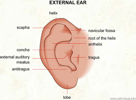
The outer ear is the most external portion of the ear. The outer ear includes the pinnae also
called auricle, the ear canal, and the very most superficial layer of the ear drum also called the
tympanic membrane. In humans, and almost all vertebrates, the only visible portion of the ear
is the outer ear. Although the word ear may properly refer to the pinna the flesh covered
cartilage appendage on either side of the head, this portion of the ear is not vital for hearing.
The complicated design of the human outer ear does help capture sound and imposes filtering
that helps distinguish the direction of the sound source, but the most important functional
aspect of the human outer ear is the ear canal itself.
Unless the canal is open, hearing will be dampened. Ear wax medical name - cerumen is
produced by glands in the skin of the outer portion of the ear canal. This outer ear canal skin
is applied to cartilage; the thinner skin of the deep canal lies on the bone of the skull. Only the
thicker cerumen-producing ear canal skin has hairs. The outer ear ends at the most
superficial layer of the tympanic membrane. The tympanic membrane is commonly called the
ear drum.
The pinna helps direct sound through the ear canal to the tympanic membrane eardrum. In
some animals with mobile pinnae like the horse, each pinna can be aimed independently to
better receive the sound. For these animals, the pinnae help localize the direction of the sound
source. Human beings localize sound within the central nervous system, by comparing
arrival-time differences and loudness from each ear, in brain circuits that are connected to
both ears.
Human outer ear and culture
Although the function of the human auricle is rudimentary in terms of hearing, the ears do
have an effect on facial appearance. In Western societies, protruding ears present in about %
of the Europeans & descendants have been considered unattractive, particularly if
asymmetric. The first surgery to reduce the projection of prominent ears was published in the
medical literature in .
The ears have also been ornamented with jewelery for thousands of years, traditionally by
piercing of the earlobe. In some cultures, ornaments are placed to stretch and enlarge the
earlobes to make them very large. Tearing of the earlobe from the weight of heavy earrings,
or from traumatic pull of an earring for example by snagging on a sweater being removed, is
fairly common. The repair of such a tear is usually not difficult.
A cosmetic surgical procedure to reduce the size or change the shape of the ear is called an
otoplasty. In the rare cases when no pinna is formed atresia, or is extremely small microtia
reconstruction the auricle is possible. Most often, a cartilage graft from another part of the
body generally, rib cartilage is used to form the matrix of the ear, and skin grafts or rotation
flaps are used to provide the covering skin. However, when babies are born without an auricle
on one or both sides, or when the auricle is very tiny, the ear canal is ordinarily either small or
absent, and the middle ear often has deformities. The initial medical intervention is aimed at
assessing the babys hearing and the condition of the ear canal, as well as the middle and inner
ear. Depending on the results of tests, reconstruction of the outer ear is done in stages, with
planning for any possible repairs of the rest of the ear.
Middle ear
The middle ear, an air-filled cavity behind the ear drum tympanic membrane, includes the
three ear bones or ossicles: the malleus or hammer, incus or anvil, and stapes or stirrup. The
opening of the Eustachian tube is also within the middle ear. The malleus has a long process
the manubrium, or handle that is attached to the mobile portion of the eardrum. The incus is
the bridge between the malleus and stapes. The stapes is the smallest named bone in the
human body. The three bones are arranged so that movement of the tympanic membrane
causes movement of the malleus, which causes movement of the incus, which causes movement
of the stapes. When the stapes footplate pushes on the oval window, it causes movement of
fluid within the cochlea a portion of the inner ear.
In humans and other land animals, the middle ear like the ear canal is normally filled with air.
Unlike the open ear canal, however, the air of the middle ear is not in direct contact with the
atmosphere outside the body. The Eustachian tube connects from the chamber of the middle
ear to the back of the pharynx. The middle ear is very much like a specialized paranasal sinus,
called the tympanic cavity; it, like the paranasal sinuses, is a hollow mucosa-lined cavity in the
skull that is ventilated through the nose. The mastoid portion of the human temporal bone,
which can be felt as a bump in the skull behind the pinna, also contains air, which is ventilated
through the middle ear.
Normally, the Eustachian tube is collapsed, but it gapes open both with swallowing and with
positive pressure. When taking off in an airplane, the surrounding air pressure goes from
higher on the ground to lower in the sky. The air in the middle ear expands as the plane gains
altitude, and pushes its way into the back of the nose and mouth. On the way down, the volume
of air in the middle ear shrinks, and a slight vacuum is produced. Active opening of the
Eustachian tube is required to equalize the pressure between the middle ear and the
surrounding atmosphere as the plane descends. The diver also experiences this change in
pressure, but with greater rates of pressure change; active opening of the Eustachian tube is
required more frequently as the diver goes deeper into higher pressure.
The arrangement of the tympanic membrane and ossicles works to efficiently couple the sound
from the opening of the ear canal to the cochlea. There are several simple mechanisms that
combine to increase the sound pressure. The first is the hydraulic principle. The surface area
of the tympanic membrane is many times that of the stapes footplate. Sound energy strikes the
tympanic membrane and is concentrated to the smaller footplate. A second mechanism is the
lever principle. The dimensions of the articulating ear ossicles lead to an increase in the force
applied to the stapes footplate compared with that applied to the malleus. A third mechanism
channels the sound pressure to one end of the cochlea, and protects the other end from being
struck by sound waves. In humans, this is called round window protection, and will be more
fully discussed in the next section.
Abnormalities such as impacted ear wax occlusion of the external ear canal, fixed or missing
ossicles, or holes in the tympanic membrane generally produce conductive hearing loss.
Conductive hearing loss may also result from middle ear inflammation causing fluid build-up
in the normally air-filled space. Tympanoplasty is the general name of the operation to repair
the middle ears tympanic membrane and ossicles. Grafts from muscle fascia are ordinarily
used to rebuild an intact ear drum. Sometimes artificial ear bones are placed to substitute for
damaged ones, or a disrupted ossicular chain is rebuilt in order to conduct sound effectively.
Inner ear Cochlea, Vestibule, and Semi-Circular Canals
The inner ear includes both the organ of hearing the cochlea and a sense organ that is attuned
to the effects of both gravity and motion labyrinth or vestibular apparatus. The balance
portion of the inner ear consists of three semi-circular canals and the vestibule. The inner ear
is encased in the hardest bone of the body. Within this ivory hard bone, there are fluid-filled
hollows. Within the cochlea are three fluid filled spaces: the tympanic canal, the vestibular
canal, and the middle canal. The eighth cranial nerve comes from the brain stem to enter the
inner ear. When sound strikes the ear drum, the movement is transferred to the footplate of
the stapes, which presses into one of the fluid-filled ducts of the cochlea. The fluid inside this
duct is moved, flowing against the receptor cells of the Organ of Corti, which fire. These
stimulate the spiral ganglion, which sends information through the auditory portion of the
eighth cranial nerve to the brain.
Hair cells are also the receptor cells involved in balance, although the hair cells of the auditory
and vestibular systems of the ear are not identical. Vestibular hair cells are stimulated by
movement of fluid in the semicircular canals and the utricle and saccule. Firing of vestibular
hair cells stimulates the Vestibular portion of the eighth cranial nerve.
Damage to the human ear
The auricle can be easily damaged. Because it is skin-covered cartilage, with only a thin
padding of connective tissue, rough handling of the ear can cause enough swelling to
jeopardize the blood-supply to its framework, the auricular cartilage. That entire cartilage
framework is fed by a thin covering membrane called the perichondrium meaning literally:
around the cartilage. Any fluid from swelling or blood from injury that collects between the
perichondrium and the underlying cartilage puts the cartilage in danger of being separated
from its supply of nutrients. If portions of the cartilage starve and die, the ear never heals
back into its normal shape. Instead, the cartilage becomes lumpy and distorted. Wrestlers Ear
is one term used to describe the result, because wrestling is one of the most common ways
such an injury occurs. Cauliflower ear is another name for the same condition, because the
thickened auricle can resemble that vegetable.
The lobule of the ear ear lobe is the one part of the human auricle that normally contains no
cartilage. Instead, it is a wedge of adipose tissue fat covered by skin. There are many normal
variations to the shape of the ear lobe, which may be small or large. Tears of the earlobe can
be generally repaired with good results. Since there is no cartilage, there is not the risk of
deformity from a blood clot or pressure injury to the ear lobe.Other injuries to the external ear
occur fairly frequently, and can leave a major deformity. Some of the more common ones
include, laceration from glass, knives, and bite injuries, avulsion injuries, cancer, frostbite,
and burns.
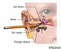
Ear canal injuries can come from firecrackers and other explosives, and mechanical trauma
from placement of foreign bodies into the ear. The ear canal is most often self-traumatized
from efforts at ear cleaning. The outer part of the ear canal rests on the flesh of the head; the
inner part rests in the opening of the bony skull called the external auditory meatus. The skin
is very different on each part. The outer skin is thick, and contains glands as well as hair
follicles. The glands make cerumen also called ear wax. The skin of the outer part moves a bit
if the pinna is pulled; it is only loosely applied to the underlying tissues. The skin of the bony
canal, on the other hand, is not only among the most delicate skin in the human body, it is
tightly applied to the underlying bone. A slender object used to blindly clean cerumen out of
the ear often results instead with the wax being pushed in, and contact with the thin skin of the
bony canal is likely to lead to laceration and bleeding.