 Return to Gross Anatomy
THE THORAX AND EARLY EMBRYOLOGY
Download a copy of this study guide
 Return to top
EARLY EMBRYONIC DEVELOPMENT
Morula: The name of the embryo at the 16 to 32 cell stage.
Blastocyst: At about day 5 after implantation.
- Cells are divided into the inner cell mass and trophoblast.
- Inner Cell Mass: The cells on the inside.
- Trophoblast: The cells surrounding the outside.
Implantation: Blastocyst travels from ampulla ------> oviduct ------> uterus to implant in the
uterus. Implantation occurs around day 5. Trophoblast forms two cell-layers.
- Syncytiotrophoblast: The cell mass at the embryonic pole (nearest the uterine wall).
- Cytotrophoblast: The cell mass jutting out from the pole. Will form the extraembryonic
membranes.
Inner Cell Mass: Form two layers of cells, epiblast and hypoblast. A Bilaminar Embryonic
Disc is formed.
- Epiblast Cells: From inner cell mass, will ultimately give rise to the three germ layers and
the entire embryo.
- Amniotic Cavity: Forms as the space between the Epiblast and cytotrophoblast..
- Hypoblast Cells: These cells are the first to migrate and eventually disintegrate.
Progression of events after implantation: Each item represents simultaneous events.
- The epiblast cells grow deeper into the wall of the uterus.
- The amniotic cavity begins to form in the epiblast layer.
- Amnioblasts differentiate from the epiblasts and migrate to the inner amniotic layer.
- Heuser's Membrane is formed from hypoblasts (interior of cytotrophoblast
layer). It will differentiate into the yolk sac.
- Trophoblastic Lacunae are openings that form in the syncytiotrophoblast.
- Maternal blood enters the lacunae.
- Extraembryonic Reticulum forms.
- Primary Yolk Sac: Epiblasts form mesoderm on the exterior of Heuser's Membrane.
- Definitive Yolk Sac: New Hypoblast cells migrate to displace Heuser's membrane,
pushing back the primary yolks sac to form the new definitive yolk sac.
- Chorionic Cavity forms in the ECM.
- The Placenta is formed from a network of villi (trophoblast cells). Villi form from
trophoblast in the following order of maturation:
- Primary Villus: Cytotrophoblast + a layer of syncytiotrophoblast grow into the
lacunae.
- Secondary Villus: Extraembryonic mesoderm grows into the primary villus.
- Tertiary Villus: Extraembryonic mesoderm gives rise to blood vessels.
Exchange of nutrients is now possible with mother.
Formation of Three Germ Layers:
- Form by migration of epiblast cells through the primitive streak.
- First Migration: Epiblasts migrate toward the midline and fold under, as they
displace the hypoblast layer. This forms the true endoderm.
- NOTE: True Endoderm comes from migration of 1st epiblast layer -- not
from the hypoblast!
- Second Migration: More epiblasts migrate to form mesoderm.
- The epiblasts that remain on the dorsal surface form the ectoderm.
Axis Determination:
- Buccopharyngeal Membrane: Forms in the future cephalic region, a tightly adhered
region through which mesoderm cannot intrude. This is the future mouth.
- Cloacal Membrane: This is the future anus.
Notochord Formation:
- Notochordal Process forms from mesoderm.
- Neurenteric canal is a transient opening between the amniotic cavity and the yolk sac.
- Notochordal plate reforms a tube now known as the notochord.
- It is the dorsal remains of the notochordal canal.
- Endoderm is once again present dorsal to it, and the secondary yolk sac is again
intact.
Concurrent Ectodermal Changes: Formation of the Neural Plate and Neural Crest Cells.
- Notochord induces ectodermal formation of the neural plate.
- As neural plate invaginates medially, forming the midline neural groove and laterally
placed neural folds.
- Neural Tube forms as the fusion of the neural folds.
- Rostral Neuropore is leading cranial edge of this folding.
- Caudal Neuropore is caudal edge of this folding.
- The Amniotic Cavity repositions itself around the embryo, in preparation for
flexion.
- Neural Crest Cells migrate outward from the neural tube to form the Dorsal Root
Ganglia and Chain Ganglia.
Other Mesodermal Structures:
- Paraxial Mesoderm: Will form the somites.
- It is directly adjacent to the neural tube.
- It participates in formation of the axial skeleton.
- Intermediate Mesoderm: Forms the urogenital system.
- Lateral to the paraxial skeleton, but medial to the lateral plate mesoderm.
- Lateral Plate Mesoderm: It is the most lateral of the three. Forms primitive gut, and
posterior and lateral body wall. It differentiates into two layers:
- Somatic Mesoderm: (Amniotic side)
- Splanchnic Mesoderm: (Yolk side)
Formation of the Intraembryonic Coelom: Forms as a division of the lateral-plate mesoderm.
- As opposed to the chorionic cavity, which is extraembryonic coelom.
- It is horseshoe-shaped, with the base of the horseshoe extending cranial.
- The horseshoe is later divided into three segments:
- The Cranial Segment (base) becomes: Pericardial Coelom.
- Left and Right Caudal portions become the: Peritoneal Coeloms.
- The junction of the U-shapes coeloms is the pericardioperitoneal canal.
- Formation of Coelom creates two mesodermal layers now:
- Somatic Mesoderm -- parietal mesoderm.
- Splanchnic Mesoderm -- visceral mesoderm.
Somite Development: Somites establish the segmental nature of the body. They differentiate into
three layers.
- Sclerotome: Forms the vertebral bone. This originates from paraxial mesoderm.
- As cells migrate outward, each sclerotome splits into inferior and superior halves.
The inferior half of one sclerotome merges with the superior half of the next
sclerotome to form the respective vertebrata.
- Two somites from each side, for a total of FOUR DIFFERENT SOMITES,
contribute to the formation of each vertebrata.
- When adjacent sclerotomes merge, the remaining notochord forms the nucleus
pulposus. The rest of the notochord degenerates.
- Dermamyotome, consisting of myotome (medial part) and dermatome (most lateral part)
Early Somite Development:
- Chorioamnionic Membrane forms, merging the chorion and amnion cavities.
- Folding (flexion) of the embryo repositions the somites from the ventral-medial position to
a dorsolateral position.
NEURAL CREST DERIVATIVES:
- Dorsal Root Ganglia
- Autonomic Ganglia
- Melanocytes
- Chromaffin Cells of Adrenal Medulla
- Enterochromaffin Cells
- Pia
- Celiac Ganglion
- Schwann Cells (but not Oligodendrocytes)
- Odontoblasts
- Parafollicular cells of Thyroid
FETAL / ADULT EQUIVALENTS:
| Fetal Structure |
Adult Structure |
| Umbilical Vein |
Ligamentum Teres Hepatis |
| Umbilical Arteries |
Medial Umbilical Ligaments |
| Ductus Arteriosus |
Ligamentum Arteriosum |
| Ductus Venosus |
Ligamentum Venosum |
| Foramen Ovale |
Fossa Ovalis |
| Allantois, Urachus |
Median Umbilical Ligament |
| Notochord |
Nucleus Pulposus |
Return to top
BONES / GENERAL REGIONS OF THORAX
STERNAL ANGLE: The junction between the Manubrium and the Sternum body.
- At the level of T4 and T5 posteriorly.
- Anteriorly, it articulates with the 2nd Costal Cartilage.
- The Bifurcation of the Trachea occurs directly posterior to the Sternal Angle.
STERNUM: The breast bone, composed of three parts.
- Manubrium: Superior portion, composed of 1st and 2nd costal cartilages.
- Sternal Body: Main middle portion. 3rd thru 7th Costal Cartilages.
- Xiphoid Process: Extending below the sternal body, not connected to any costal
cartilages.
CLAVICLE: Collar bone. It covers the 1st rib, so that the 1st rib cannot be palpated.
STERNOCOSTAL JOINTS: Joint between the sternum and each costal cartilage.
COSTOCHONDRAL JOINTS: Joint between each costal cartilage and respective rib.
COSTOVERTEBRAL JOINTS: Joint between each rib and vertebrata, joined at the transverse
process of the vertebrata and the articular facet of the rib (at the tubercle).
SUPERIOR MEDIASTINUM: Defined as the region of the thorax above the sternal angle
(above T4-T5).
- Contains the Aortic Arches and the Thymus.
INFERIOR MEDIASTINUM: Defined as the region of the thorax below the Sternal Angle.
Divided into Anterior, Middle, and Posterior Mediastinum.
- Middle Mediastinum: The pericardial sac, holding the heart.
- Posterior Mediastinum: The esophagus and trachea.
- Anterior Mediastinum: Not much is there.
THE MID-AXIAL LINE: The side of the thorax. The line running from the armpits to the hips.
THE SUPERIOR THORACIC APERTURE: Surrounded by the clavicle, the superior border of
the thorax, above which is the base of the neck.
THE BORDERS OF THE THORAX:
- Superior Border: Superior Thoracic Aperture.
- Inferior Border: Diaphragm.
- Anterior Border: The sternum.
- Posterior Border: Thoracic Vertebrata, T1-T12
- Lateral Border: Mid-Axial Line.
THE RIBS:
- They articulate with the Thoracic Vertebrata. As they come anteriorly, they move
inferiorly and then stop. Then, the Costal Cartilage comes back a little superiorly.
- That means, in the back along the spinal column, the level is up higher than the
same level in the front along the sternum.
- True Ribs: T1-T7. They have their own Costal Cartilage on the anterior side.
- False Ribs: T8-T10. They all share one common costal cartilage (which mends into each
other)
- Floating Ribs: T11 and T12. They have no Costal Cartilage.
- 1st Rib:
- It has no angle.
- It is the shortest and flattest of all ribs.
- It has grooves for the subclavian veins and arteries.
COMPONENTS OF A RIB:
- Head: Connected to the Vertebrata.
- Neck
- Tubercle: Contains the articular facet, which articulates with the transverse process of
the vertebrata.
- Angle
- Shaft, containing the Costal Groove.
- Costochondral Joint: The joint where bone ends and cartilage begins. The bone extends
anteroinferiorly, whereas the cartilage extends back posterosuperiorly.
- Costal Groove: Groove for intercostal arteries, veins, and nerves, on inferior aspect.
COMPONENTS OF THORACIC VERTEBRATA:
- The body is the anterior portion of Thoracic Vertebrae.
- The Spinous Process articulates posteriorly.
- The Transverse Process articulates laterally and contains the facet for each rib, at the
costovertebral joint.
- The Pedicle is the short, thicker section that connects the transverse process to the body.
- The Lamina is the longer, thinner section that connects each transverse process to the
spinous process.
Return to top
THE BREAST
BREASTS / MAMMARY GLANDS:
- Cooper's Ligaments: Suspensory Ligaments, or connective tissue that connects the skin
to the underlying fat.
- Lactiferous Sinuses: Place where the milk is stored.
- Deep to the areola.
- The dilated portion of the lactiferous ducts.
- Lactiferous Ducts: The ducts into which milk is secreted. Directly deep to the nipple.
- Areola: Darkened region around nipple. Appears lighter in women who have not born a
child.
- Contains sebaceous glands which secrete protective substance (not milk) during
pregnancy.
- Mammary Glands: Lobules of glandular tissue which arise from the lactiferous ducts.
Any deep tissue that is not fatty is the glandular stuff.
BREAST CARCINOMA: Lymphatic drainage of the breast explains the danger of breast
cancer.
- Principle Axillary Lymph Node (in region of armpit) takes about 75% of lymph
drainage from breast.
- Path of this node: Axillary Lymph Node ------> Subclavian Trunk ------> Jugular
Vein. Hence cancer can easily metastasize to all parts of the body from the breast.
Return to top
MUSCULATURE OF THE THORAX
Muscles Associated with Movement of Arm and Neck: These muscles are relevant to movement
of upper limbs.
- Pectoralis Major: Most anterior portion.
- Pectoralis Minor: Deep to pectoralis majora.
- Scalenus Muscles
Muscles of the Thorax Proper: All of these muscles are associated with respiration.
- Serratus Posterior (Outer Muscles): Originate from the vertebrata and insert on the ribs
on the posterior.
- Serratus Posterior Superior: Insert on superior ribs posteriorly. When they
contract, they raise the ribs.
- Serratus Posterior Inferior: Insert on inferior ribs posteriorly. When they
contract, they lower the ribs.
- Inserts on the anterior surface (body) of the vertebrata and the anterior
surface of the ribs.
- Levatores Costarum (Outer Muscles): Small muscles that pass all along the vertebral
column. Raise the ribs upon contraction.
- Intercostal Muscles: Inner Muscles that pass between the ribs.
- External Intercostals:
- Pass from Lateral to Medial
- Originate at Vertebrata and travels along intercostal space.
- Stops before it gets to the sternum.
- External Intercostal Membrane: Connects the muscle to the sternum on
the anterior side.
- Internal Intercostals: Deep to the external intercostals.
- Run from medial to lateral, crossing the external intercostals.
- Start at the sternum, pass laterally and posteriorly, and stop around the
mid-axial line.
- The Internal Intercostal Membrane connects the muscles to the
vertebrata at that point.
- Innermost Intercostals: Start at the angle of the ribs, and move anteriorly and
stop before the sternum.
- They only cover the lateral region of the thorax.
- LAB: How to tell the intercostals:
- The anterior aspect of thoracic wall: you can see the pectorals (shallow) and the
internal intercostals (deep).
- The lateral aspect of the thoracic wall: you can see mostly the innermost
intercostals.
- The posterior aspect of the thoracic wall: you can see the external intercostals and
the serratus posteriors.
- Transversus Thoracis: Can be seen from innermost aspect of thorax. Connected to
innermost intercostals through a membrane.
- Subcostals: Located on the posterior aspect, internally. Slips of muscle that pass
between the posterior ribs.
Return to top
NERVE AND BLOOD SUPPLY TO THE INTERCOSTALS
V.A.N: Going from superior to inferior, the order of each intercostal space is vein, artery, nerve.
Origin of Intercostal Nerves:
- Mixed Nerve: Sensory Nerves come from the Dorsal Root of the spinal chord. Motor
nerves come from the ventral root of the spinal chord. These two merge together to
become one nerve.
- The nerve also received fibers from the Sympathetic Chain Ganglia, the
sympathetic part of the ANS.
- The mixed nerve branches into Doral Ramus and Ventral Ramus.
- Dorsal Ramus: Innervates the back muscles.
- Ventral Ramus: Innervates the intercostal muscles.
Arterial Blood Supply to the intercostal muscles: Intercostal Arteries. The vasculature branches
as follows: Subclavian Artery ------> Internal Thoracic Artery ------> Intercostal Artery.
- Internal Thoracic Artery: Travels Superoinferiorly, lateral to the sternum, deep to the
costal cartilages.
- Costal Groove: Intercostal arteries, veins, and nerves run along this path, on the inferior
aspect of the ribs.
Clinical Application: To place a needle through the ribs, it should be placed in the center of the
intercostal space, because the vessels and nerves (VAN) run along each rib on the costal groove.
Return to top
EMBRYONIC PARTITIONING OF BODY CAVITIES
Embryonic Period: Five-week interval of fourth through eighth weeks, when primary
differentiation occurs.
Fetal Period: Eight week to term, when primarily growth occurs, secondary differentiation, and
some primary differentiation (such as urogenital system).
EMBRYONIC FLEXION: Folding of the embryo.
- Transverse Folding: Right and left lateral-medial folding.
- Splanchnopleure of the yolk sac will merge to form the midgut in folding.
- The intraembryonic coelom is the primitive peritoneum. It will differentiate into
the mesothelium, and blood vessels will pass through it as it forms the serous
peritoneal membrane.
- Longitudinal Folding:
- HEAD FOLD: Before the folding occurs, the presumptive heart tissue is Cranial
to the primitive mouth -- oropharyngeal membrane. After folding, this is
reversed. The membrane is the axis for the 180 turn of:
- Septum Transversum turns 180 along the oropharyngeal axis.
- Pericardial Coelom turns 180.
- Cardiogenic Tissue turns 180.
- During Head-folding, these structures turn so that they are now caudal to
the oropharyngeal membrane.
- TAIL FOLD: Longitudinal folding here takes place about the axis of the cloacal
membrane.
- Part of yolk sac is incorporated into embryo to make hindgut.
- AFTER FOLDING
- The pericardial coelom moved from dorsal to ventral, relative to the developing
heart tissue.
- Septum Transversum became cranial (rather than caudal) to the developing heart
tissue.
- Primitive Streak was cranial to the cloacal membrane. Now it is caudal.
- The connecting stalk is pushed ventral-medially and becomes the umbilical cord.
- SUMMARY OF FOLDING EVENTS AND CONSEQUENCES
- Primitive Gut was formed from the dorsal regions of the yolk sac.
- The embryo changes in shape from a "trilaminar disc" to a crescent C-Shape.
- Lateral and Ventral body walls are formed.
- Primitive body cavities are subdivided -- pericardial and peritoneal.
- Future mouth is defined.
- Umbilical chord is defined.
Septum Transversum: The posterior border of the developing heart. It will develop into the
diaphragm.
- It is the primitive central tendon of the adult diaphragm.
- It rotates around the foregut like a blanket, hence giving rise to the esophageal hiatus.
Pericardioperitoneal Canals: Two canals in the embryo. Between the two canals is the
foregut.
- The Thoracic Cavity is formed as a result of forming the diaphragm.
- Each canal contains a pleuroperitoneal membrane.
- An ingrowth of these membranes closes off the pericardioperitoneal canals.
Muscle wall moves in with it, eventually forming the diaphragm.
- The Central Tendon of the diaphragm is a remnant of the septum transversum.
- Birth Defect: The GI-Tract may expand into the Thoracic cavity if the diaphragm
does not fully form (i.e. the separation between the cavities is incomplete). A
congenital Hiatal Hernia
Innervation of Diaphragm
- The Phrenic Nerve innervates the diaphragm.
- Phrenic nerve originates at C3-C5.
- During development the diaphragm is displaced inferiorly. It drags the phrenic nerve
along with it.
Referred Pain: Chest pain, lung pain, or pain in the diaphragm may be felt in the left shoulder.
This is because the sensory origins of the phrenic nerve (C3-C5) also innervate the left shoulder
region. When the pain is displaced in this manner, it is called referred pain.
Lung Development:
- Primitive Pharynx: The floor of the foregut, where the lungs develop.
- Laryngeal-Tracheal Groove: A groove in the primitive pharynx.
- The lung bud originates from this groove. This tube-like structure bifurcates to
form the bronchi, and continues to bifurcate to form the entire alveolar system.
Pleural Cavity Development: Two membranes structures. Imagine them as balloons. Stick your
fist into the balloons. The fist is the lung, from the lung root, and the balloons are the pleural
membranes. Hence the lungs are surrounded by a double-membrane system.
Return to top
THE LUNGS
Respiration: The thoracic cavity is enlarged, decreasing pressure, resulting in air being sucked
into the lungs. Negative pressure breathing.
- Expansion of rib-cage.
- Ribs 1-7 rotate from respiration. They undergo the pail-handle effect. As they are
raised, they increase volume in two dimensions:
- Anterior-posterior dimension.
- Medial-Lateral dimension (because they are bowed)
- Ribs 9-12 bow out laterally, increasing volume in both dimensions described
above.
- Vertical Increase (superior-inferior) is the most significant: It results from contraction of
the diaphragm, displacing it inferiorly.
PLEURAL CAVITY: The two separate cavities in which the lungs are contained.
- Visceral Pleura: The inner membrane of the pleural cavity, or the membrane immediately
surrounding the lung.
- Parietal Pleura: The outer membrane of the pleural cavity.
- The Costal Pleura: That portion of the parietal pleura bordering the rib-cage.
- The Mediastinal Pleura: That portion of the parietal pleura bordering the
mediastinum.
- The Diaphragmatic Pleura: That portion of the parietal pleura bordering the
diaphragm.
Costodiaphragmatic Recess: The inferior cavity between the bottom (base) of the lung and the
diaphragm.
- Whenever fluid accumulates in the pleural cavity (pleuritis), this is a good place to put a
drain tube.
Costomediastinal Recess: The recess created by the junctions (reflections) between the
mediastinal pleura and costal pleura.
- On the anterosuperior portion of both lungs.
- The left costomediastinal recess is larger than the right, due to the cardiac notch -- the
impression left on the left lung from the heart.
Borders of the Lungs:
- Inferior Border: The 6th rib (anteriorly), and T-10 (posteriorly)
- Lateral Border: The mid-axillary line.
- Superior Border: The lungs extend through the Thoracic Aperture into the base of the
neck.
Apex of the Lung: The top of the lung, just superior and deep to the clavicle.
- The Subclavian Artery crosses the apex of the lung.
Base of the Lung: The concave inferior border of the lung, having the same curvature as the
convex surface of the diaphragm.
Root of the Lung: The center of the lung at its attachment point, wherein lies the hilus.
Hilum of the Lung: Where the root is attached. Contains all the arteries, vessels, nerves, and
lymph vessels of the lung. The hilum contains the following.
- Pulmonary Arteries.
- Middle and Lower Lobe Bronchi
- Lymph Nodes
- Superior and Inferior Pulmonary Veins
DEPRESSIONS IN LUNGS: Both lungs have the following depressions in the superior region of
the lungs
- Brachiocephalic vein.
- Subclavian artery
- Trachea (may be seen on right lung more than left, as trachea is more toward the right
side).
- Esophagus
THE RIGHT LUNG: The larger of the two lungs. The heart extends into the left lung (at the
cardiac notch). The right lung has no cardiac notch. Has three lobes.
- Horizontal Fissure: separates the Upper Lobe from the Middle Lobe..
- Oblique Fissure: Separate both of the other lobes from the Lower Lobe.
- Hilus: The pulmonary artery is between the bronchi and pulmonary veins.
- Bronchi are most posterior.
- Pulmonary artery is between bronchus and pulmonary vein.
- Arteries and bronchi are in relatively the same superior/inferior plane.
- Epartial Bronchus: The bronchus closely associated with the superior pulmonary
artery in the right lung. It ventilates the upper lobe.
- Depressions: Right Lung has depression for superior vena cava.
THE LEFT LUNG: The smaller of the two lungs, containing a recess for the Cardiac Notch.
- Oblique Fissure: Separates the Upper Lobe and Lower Lobe. There is no middle lobe.
- Hilus:
- Bronchi are still most posterior. The left bronchus is posterior to the pulmonary
arteries.
- Pulmonary artery is most superior.
- Depressions: Left Lung has big groove for Aortic Arch.
- Cardiac Impression: In addition to the cardiac notch, the left lung has an impression
where the heart is, anterior to the hilus.
Bronchopulmonary Segments: An arterial supply to the lungs develops along with the bronchi,
resulting in different parts of the lungs being perfused by different blood supplies.
Return to top
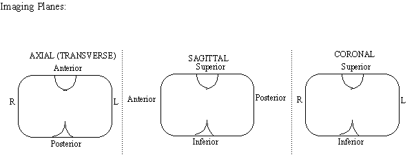
FUNDAMENTALS OF RADIOLOGIC ANATOMY
Requirements for Medical Imaging:
- An energy source (light or sound)
- An object (the patient)
- A detector
Conventional Radiography: X-Rays
- When X-Rays strike photographic film (silver halide granules), they blacken the film. X-rays that get through indicate the absence of absorption, and hence black is low density.
- Conventional Objects and their corresponding absorptions:
- Air: Black
- Fat: Grey
- Soft Tissue: Various shades of grey
- Bone: White
- Barium or Iodine can be given to visualize soft tissue that was otherwise not visible.
- Example: Lower GI can be given by giving a Barium enema, making the Colon visible.
- Arteriogram: Injecting visible substances into arteries for purposes of imaging.
Sectional Imaging: MRIs, CTs, Sonograms. Multiple pictures (Sections) given in an image
plane.
 Computerized Topography: CT-Scan
- Computer generated reconstruction of sectional images.
- X-Rays are passed through at many different angles, and a 3D-image is generated as a
result by a computer.
Magnetic Resonance Imaging: MRI
- Monitors the magnetic moments of H-nuclei in tissues.
- Uses a very strong magnet and radio waves.
- Imaging is the opposite as X-Rays:
- Cortical bone and dense tissue appears black.
- Air + Soft Tissue + Fat appears white.
- MRI cannot be used if patient has magnetic metal anywhere in body (such as tooth filling).
Ultrasound Imaging: Variable pattern of bright dots and dashes on a black screen.
- There are theoretical risks to the mechanical waves, but no known risks.
Return to top
EMBRYOLOGIC DEVELOPMENT OF THE HEART
AORTIC ARCH DERIVATIVES:
| Aortic Arch |
Adult Structures |
| 1st Aortic Arch |
Part of Maxillary Artery |
| 2nd Aortic
Arch |
Stapedial Artery
Hyoid Artery |
| 3rd Aortic Arch |
Common Carotid Artery
Proximal part of Internal Carotid Artery |
| 4th Arotic Arch |
Left Arch: Gives rise to Aortic Arch
Right Arch: Proximal part of Right
Subclavin Artery |
| 5th Aortic Arch |
Degenerates |
| 6th Aortic Arch |
Both Arches: Proximal part of pulmonary
arteries
Left Arch only: Ductus Arteriosus |
EMBRYOLOGIC HEART DERIVATIVES:
| Embryonic Structure |
Adult Structure |
| Bulbus Cordis |
Right Ventricle and Aortic Outflow Track |
| Primitive Ventricle |
Left Ventricle |
| Truncus Arteriosus |
Ascending Aorta, Pulmonary Trunk |
| Primitive Atria |
Auricular Appendages |
| Sinus Venosus |
Left Horn: Coronary Sinus
Right Horn: Smooth part of the Right Atrium |
| Right Common and Anterior Cardinal Veins |
Superior Vena Cava |
FUSION of the HEART TUBES: Day 20
- From Cranial to Caudal end, the heart is composed of the following after fusion of the
heart tubes:
- Truncus Arteriosus: Will lead to six pairs of aortic arches later in development.
Truncus Arteriosus itself eventually becomes the ascending Aorta.
- Bulbus Cordis: Eventually forms the right ventricle and Aortic Outflow tract.
- Primitive Ventricle: Eventually forms the adult left ventricle.
- Primitive Atrium: Eventually becomes the auricular appendages of the adult atria.
- Sinus Venosus: Will be enveloped by the septum transversum and hence become
a part of the developing diaphragm.
- At birth, Right horn of the sinus venosus will merge with Right Atrium.
This merging results in the smooth tissue called the Sinus Venarum.
- Crista Terminalis: Demarcation point where the right horn was
incorporated into the Right Atrium.
- At birth, Left horn of the Sinus Venosus will become the Coronary Sinus.
- INVERSION: The atrium becomes caudal to the ventricle by an inversion of the heart
tube.
- The ventricle goes more posterior as growth continues.
- The atrium goes more superior as growth continues.
- PARTITIONING of the ATRIA:
- Atrioventricular Canal: An ingrowth of membrane called the endocardial
cushion is at the bottom of the canal.
- First, the Septum Primum grows toward the endocardial cushion, forming two
chambers.
- Then a hole in the new wall develops, called the foramen secundum.
- Then a second septal wall starts to form (in future Right Atrium), called the
Septum Secundum. This septum grows covering the first septal wall.
- Foramen Ovale: The Septum Primum then degenerates, leaving enough tissue to
leave a hole between the first and second septa. The Foramen Ovale Functions as
a valve in the primitive embryo.
- FETAL CIRCULATION: The embryo does not have functional lungs, and the pulmonary
circulation is thus bypassed.
- Embryonic blood flow: Right Atrium ------> (Tricuspid Valve) ------> Right
Ventricle ------> Foramen Ovale ------> Left Ventricle ------> Aorta.
- Partitioning of the Ventricles: The Interventricular Septum grows inward, similar to the
growth of the Septum Secundum.
TETRALOGY OF FALLOT: Developmental error where the Pulmonary Trunk fails to
develop fully, and instead the cells are allotted to the Aorta, resulting in an overriding Aorta. The
result is that blood bypasses the Pulmonary Trunk and never gets oxygenated by the lungs.
- Left Ventricular Hypertrophy: Left Ventricular Wall becomes enlarged as the Left
Ventricle must work harder to accommodate a higher flow of blood.
- Blue Baby: Hypoxia results from insufficient oxygenation of blood.
Return to top
PERICARDIUM AND POSITIONING OF THE HEART
Base of the Heart: The superior border of the heart, where the great vessels converge. On the
anterior surface of the heart, near the Right Atrium.
Apex of the Heart: The inferior border of the heart, at the anterior portion of the Left Ventricle.
Fibrous Pericardium: The outer membrane (parietal membrane) of the Pericardial Sac.
- Sternal Pericardial Ligaments connect the fibrous pericardium to the sternum. These
ligaments help hold the heart in place.
- The Phrenic Nerve is embedded in the fibrous pericardium.
Serous Pericardium: The inner membrane (visceral membrane) of the Pericardial Sac.
- It secretes a fluid into the pericardium to lubricate the heart when it is beating.
- Membrane plus fluid becomes a whole surface layer called the Epicardium.
Reflection Points: Wherever there is a vessel, the Serous Membrane will reflect into the Fibrous
Pericardium.
Right Border of the Heart: 1 cm to the right of the 3rd costal cartilage.
Left Border of the Heart: 2cm to the left of the 2nd costal cartilage.
Right Inferior Border of the Heart: 1 cm to the right of the 6th costal cartilage.
Left Inferior Border (Apex) of the Heart: 6cm to the left of the 5th Intercostal Space.
Transverse Pericardial Sinus: The hole created between the Aorta and Pulmonary Trunk, and
the Fibrous Pericardium, on the Anterior Surface of the Heart at its base. Fluid can accumulate
there.
Oblique Pericardial Sinus: The hole created by the four Pulmonary Veins, and fibrous
pericardium, on the Posterior Surface of the heart near the Left Atrium. Fluid can accumulate
there.
External Location of Heart Valves: 3, 3.5, 4, 4.5 Rule. This is not the best place to hear the
valves, but simply where they are located.
- 3rd Costal Cartilage: Location of Pulmonary Valve.
- (Right Ventricle <====> Pulmonary Trunk)
- 3rd Intercostal Space: Location of Aortic Valve.
- (Left Ventricle <====> Aorta)
- 4th Costal Cartilage: Location of Mitral Valve
- (Left Atrium <====> Left Ventricle)
- 4th Intercostal Space: Location of Tricuspid Valve.
- (Right Atrium <====> Right Ventricle)
Valve Sounds: The sounds are reflected to other places externally. The best place to hear each
valve is as follows:
- Pulmonary Valve: 2nd Sternocostal Joint
- Aortic Valve: Position of the right 2nd Costal Cartilage.
- Bicuspid Valve: Best heard at the Apex of the Heart: 8 cm to the left of the 5th
Intercostal space.
- Tricuspid Valve: Best heard in the lower left quadrant of the sternum.
INNERVATION OF THE HEART:
- Sino-Atrial (SA) Node: Located on the anterior surface of the Superior Vena Cava.
- Innervated by both sympathetic and parasympathetic fibers.
- Nervous impulse originates at the SA Node.
- Atrio-Ventricular Node: Located on the right side of the intra-atrial septal wall.
- Moderator Band: Visible in the Right Ventricle, represent fibers of the AV-Node
that go into the Right Ventricle.
Return to top
THE HEART
EXTERNAL ANATOMY
- Sulcus: Depressions on the anterior surface of the heart, used as demarcations for
external anatomy. They may be hard to see if fat is present.
- Interventricular Sulcus: The demarcation between the left and right ventricles.
The Anterior Interventricular Artery is often embedded in this sulcus.
- Coronary Sulcus (Aorticoventricular Sulcus): The border between the Right
Atrium and Aorta. The Right Coronary Artery often travels along this sulcus.
- Coronary Sinus: The Great Coronary Vein empties into the Coronary Sinus, which in
turn Empties into the Pulmonary Artery into the Right Atrium.
- The Coronary Sinus is located deep to the great vein, on the posterior wall of the
Right Atrium.
- Coronary Arteries: Originate from the right and left sides of the Ascending Aorta.
There are many variations, but common theme is below.
- Right Coronary Artery: Travels along the Atrioventricular Sulcus (Coronary
Sulcus). Then it travels posteriorly around the heart and anastomoses (joins) with
the left Coronary Artery on the posterior side.
- Left Coronary Artery: Is itself very short. It bifurcates into two more arteries:
- Circumflex Branch: Goes posteriorly and joins with the Right Coronary
Artery.
- Anterior Interventricular Branch: Travels along the Interventricular
Sulcus on the anterior side.
- Cardiac Veins: Most Cardiac veins empty into the Coronary Sinus, but not all.
- Great Cardiac Vein: Passes along the Interventricular Sulcus, with the Anterior
Interventricular Coronary Artery. It empties anteriorly into the Coronary Sinus.
- Middle Cardiac Vein: Travels with the posterior (right) interventricular
coronary artery and empties into the Coronary Sinus posteriorly.
- Anterior Cardiac Vein: An exception. It empties right into the wall of the Right
Atrium.
- Thebesian Veins: Small venous structures within the heart tissue. Only
histological structures and not visible in lab.
- Vessels of the Heart:
- Anterior Aspect, from Right to Left: Superior Vena Cava, Aorta, Pulmonary
Trunk.
- Posterior Aspect: Four Pulmonary Veins, the Inferior Vena Cava.
- Right Auricle: The primitive Right Atrium.
- Left Auricle: The primitive Left Atrium.
Vessels of the Heart: Blood Flow
- Right Atrium: Receive blood from Superior and Inferior Vena Cavae. Deliver through
Tricuspid Valve.
- Right Ventricle: Deliver blood through the Pulmonary Trunk.
- Left Atrium: Receive blood from the four Pulmonary Veins. Deliver through bicuspid
valve.
- Left Ventricle: Out the Aorta.
THE RIGHT ATRIUM:
- Musculi Pectinate: A rough area on the superior inner wall of the Right Atrium, left over
from the embryonic heart.
- Sinus Venarum: A smooth area in the Right Ventricle, remaining from the Right Horn of
the embryonic Sinus Venosus.
- Cristae Terminalis: Ridge on superior anterior border, demarcating the embryonic heart
(auricle) from the adult heart. It is at the border of the Right Auricle.
- Fossae Ovalis: Depression in the Septal wall, remaining from the embryonic Foramen
Ovale.
- Membranous Septum: A membranous remnant of the embryonic heart, smaller than the
Fossae Ovalis. It may not form, resulting in a "hole" in the septal wall of the heart.
THE RIGHT VENTRICLE:
- Chordae Tendineae: The ligaments that connect the tricuspid cusps to the Papillary
muscles, allowing them to open when the papillary muscles are contracted.
- Papillary Muscles: The muscles which control the cusps of the tricuspid valve. They are
contracted before the contraction of cardiac muscle, to close the valves, to prevent
backflow of blood into the Right Atrium.
- Trabeculae Carnea: The muscles of the Right Ventricular Wall.
- Conus Arteriosus: Superior left surface of the right ventricle, smooth.
- Tricuspid Valve: Connected to the papillary muscles via the chordae tendineae.
Composed of three cusps:
- Anterior cusp
- Posterior cusp
- Septal cusp
- Pulmonary Valve: Composed of three semilunar cusps. The valve which controls
backflow back into the right ventricle from the pulmonary trunk.
THE LEFT ATRIUM:
- Fossa Ovalis Should be visible on the septal wall.
- Bicuspid (Mitral) Valve should also be visible.
THE LEFT VENTRICLE: The largest of the chambers, with the thickest walls. The Posterior
part of the hart. Generally similar to Left Ventricle.
- Mitral Valve: Has Posterior and Anterior Cusps, and Chordae Tendineae and Papillary
Muscles, like the Right Ventricle.
- Aortic Valve: Composed of three semilunar valves: right, left, posterior.
- Coronary Sinuses: Just superior to Aortic Valve, openings for the Left and Right
Coronary Arteries.
Return to top
SUPERIOR MEDIASTINUM / NERVES AND ARTERIES OF MEDIASTINUM
Thymus: Anterior most structure in posterior mediastinum. Atrophied in adults but prominent in
children.
Ligamentum Arteriosum: Connective tissue connecting the Aorta to the Pulmonary Trunk,
helping to hold both structures in place. Left side of heart, superior to the Pulmonary Trunk.
- Developmentally, it is the former Ductus Arteriosus (Left 6th Aortic Arch) in the
embryonic heart.
The Great Veins: Anterior to the great arteries, in the superior mediastinum.
- Superior Vena Cava: Formed by the combining of the right brachiocephalic vein and left
brachiocephalic vein.
- Combination of Right and Left Brachiocephalic Vein occurs at the articulation of
the 1st rib.
- Right Brachiocephalic Vein: Right branch of Superior Vena Cava.
- Right Internal Jugular Vein: Converges on the Right Brachiocephalic Vein.
- Right Subclavian Vein: Converges on the Right Brachiocephalic Vein, and runs
anterior to the Subclavian Artery.
- Left Brachiocephalic Vein: Left branch of Superior Vena Cava.
- Left Internal Jugular Vein: Converges into the Left Brachiocephalic just lateral
to the Common Carotid Artery.
- Left Subclavian Vein: Converges into the Left Brachiocephalic Vein and runs
anterior to the Subclavian Artery.
The Great Arteries: Posterior to the great veins.
- Aorta: Ascending Aorta curves posteriorly and a bit to the left. It has three branches:
- Brachiocephalic Trunk: Right-most branch off of the Aortic Arch.
- Right Subclavian Artery: Branches off the brachiocephalic trunk.
- Left Common Carotid Artery: The center of the three branches off the Aortic
Arch.
- Left Subclavian Artery: The left-most branch off the Aortic Arch.
Internal Thoracic Arteries: Continue off of each of the Subclavian Arteries. They move down
the Thorax into the abdomen, lateral to the Sternum.
Phrenic Nerves: Both originate from C3, C4, C5. Both Phrenic Nerves are more lateral than the
Vagus nerves.
- Right Phrenic Nerve:
- Runs laterally along the Right Internal Jugular Vein.
- Continues lateral to the Superior Vena Cava.
- Then rungs along the Fibrous Pericardium.
- Finally into the diaphragm.
- Left Phrenic Nerve:
- Rungs laterally along the Left Internal Jugular Vein.
- Anterior to the Arch of the Aorta
- Then along the Fibrous Pericardium
- Into the diaphragm.
- Both Phrenic Nerves:
- They run anterior to the roots of the lungs
Vagus Nerves: Both Vagus Nerves are more medial than the Phrenic Nerves.
- Left Vagus Nerve:
- Runs lateral to the Aortic Arch.
- Gives off a branch for the Left Recurrent Pharyngeal Nerve.
- Runs anterior to the subclavian, then posterior to vena cava and brachiocephalic
veins.
- Continues medially and runs toward the diaphragm lateral to the Esophagus. In
the thorax, it tends to go to the anterior portion of the esophagus.
- Right Vagus Nerve:
- Runs lateral to the Right Common Carotid Artery (medial to Phrenic Nerve).
- Gives off a branch for the Right Recurrent Laryngeal Nerve.
- In the thorax, it tends to go to the posterior part of the esophagus.
- Both Vagus Nerves:
- Run posterior to the roots of the lungs.
- Both give off branches for the Pulmonary Plexus, Cardiac Plexus, and
Eosphageal Plexus.
- Right and left fibers mix to form the eosphageal plexus.
Recurrent Laryngeal Nerves: Both branch off the Vagus nerves and go back superiorly toward
the larynx.
- Left Recurrent Laryngeal Nerve: Off of the Left Vagus.
- Runs back up, lateral to the Trachea, into the Larynx.
- Is different in position than the Right Laryngeal, due to the degeneration of the
right 6th Aortic Arch (see below),
- Right Recurrent Laryngeal Nerve: Off of the right vagus.
- Passes back up posterior to the Right Subclavian.
- Runs back up, lateral to the Trachea, to the Larynx.
- CLINICAL: Carcinoma of the Lungs can affect the Recurrent Pharyngeals, causing a
hoarse voice. They must be watched in surgery.
Pericardiacophrenic Artery and Vein: Run on either side of the Phrenic nerve all along its path
in the Thorax.
Cardiac Plexus: Grouping of Vagal nerves innervating the heart.
Pulmonary Plexus: Grouping of Vagal nerves innervating the lungs.
Aortic Arches: The development of the Aortic Arches effected the positioning of the Right and
Left Recurrent Laryngeal nerves. They are not symmetric with respect to each other.
- There are six Aortic Arches. The 1st, 2nd, and 5th degenerate, while the 3rd, 4th, and 6th
remain behind.
- Initially the right and left laryngeal nerves pass inferior to the 6th Aortic arch, on both
sides.
- Right 6th Aortic Arch degenerates! Consequently, the right 6th Laryngeal Nerve catches
onto the 4th arch on the right side, which subsequently becomes the Right Subclavian
Artery.
- The Left 6th Aortic Arch sticks around in the embryonic heart, as the Ductus Arteriosus,
a failsafe shunt in case the foramen ovale passage fails.
- After birth the Ductus Arteriosus becomes the Ligamentum Arteriosum.
Bifurcation of the Trachea:
- Carina: The cartilage that sticks out at the bifurcation.
- Right Bronchus: Fatter and shorter than the left bronchus. It branches off at a straighter
angle, so things tend to lodge in the right Bronchus as opposed to the left.
- Left Bronchus: Branches off at a sharper angle than the right bronchus.\
Esophagus: Displaced to the right in the Thoracic Cavity. It returns to the left after it crosses
the diaphragm and goes into the abdomen.
- Eosphageal Plexus: Formed of Vagus nerve, innervates the esophagus.
- When the plexus enters the abdomen, it coalesces back into two Vagus Nerves.
Thoracic Duct: The largest lymph vessel in the body.
- To the right of the Thoracic Vertebrata, posterior to the esophagus.
- It empties into Left Brachiocephalic and Internal Jugular veins.
- This duct drains the lower half of the body and the left side of the upper body.
- Right Subclavian Lymphatic Duct empties the right half of the upper body.
Azygos Vein: Posterior to Esophagus, to the right of the Thoracic Duct.
- It is an alternate route for the return of venous blood to the heart, rather than through the
inferior vena cava.
- Intercostal veins empty into the azygos system, from both left and right (via Hemiazygous
system) sides.
- Azygos vein connects to the inferior vena cava at the level of the kidneys.
Hemiazygos Vein System: Posterior to the descending Aorta on the left side of the vertebral
column.
- It drains the left intercostal veins.
- It drains into the Azygos Vein.
Sympathetic Chain Ganglia: Lateral to the spinal column, from Cervical to Sacral.
- Intercostal Nerves: Come off of the Sympathetic Chain Ganglia in the thorax.
- Splanchnic Nerves:
- Greater Splanchnic Nerve: Comes off of the sympathetic chain at T5 to T9.
- Lesser Splanchnic Nerve: Comes off of the sympathetic chain at T10 and T11.
- Least Splanchnic Nerve: Comes off of the sympathetic chain at T12.
- Autonomic Nervous System: Location of cell bodies
- Sympathetic Nerves: The cell body is close to the spinal column. The synapse
between pre-ganglionic and post-ganglionic nerves occurs in the Chain Ganglia
near the spinal chord.
- Parasympathetic Nerves: The cell body is close to the target organ. The
synapse occurs near the target organ, with short axons innervating the target.
- Rami Communicans: The junctions where the pre-ganglionic nerves synapse with the
post-ganglionic nerves, in the sympathetic chain ganglia.
Return to top
TETRALOGY OF FALLOT LECTURE
Auscultation of Heart, Clinical Sign: The second heart sound is singular, whereas it should be
double. There should be one sound for the pulmonary valve and one for the aortic valve. In this
case there is none for the pulmonary valve.
Cyanosis: Blue-baby. The child stays blue, despite the fact that arterial blood gas breathing is
100%! The oxygen taken in lungs never gets perfused by (enough) blood.
Tetralogy of Fallot: Classic symptoms
- Four classic symptoms
- Pulmonary Trunk Stenosis
- Right Ventricular Hypertrophy (from right ventricle having to work too hard)
- Ventricular Septal Defect
- Overriding Aorta (Aorta unbent)
- Other common symptoms:
- Atrial septal defect (hole in the atrial septum)
- Right side of the Aortic Arch may be abnormal
- Coronary artery abnormalities
- Blood Flow
- Normal: Blood enters right atrium ------> right ventricle ------> pulmonary
trunk ------> lungs where it gets oxygenated
- Tetralogy: Blood enters right atrium ------> right ventricle ------> Aorta, due
to pulmonary stenosis and a hole in the ventricular septum.
Embryology of the Heart -- Tetralogy
- The problem occurs during the development of the cardiac outflow tract (the
conus/truncus area).
- Development of this area occurs during the first 8 weeks of embryonic life.
- Heart defects resulting from abnormal conus/truncus development are grouped together as
"conotruncal defects". Tetralogy of Fallot is one type of conotruncal defect.
- Other heart defects in this category include: Truncus Arteriosus, Transposition of the
Great Vessels and Double Outlet Right Ventricle.
- The developmental error that results in tetralogy of Fallot is probably abnormal
(asymmetrical) division of the embryonic truncus arteriosus.
- Some of the structural tissue of the conotruncus is derived from the neural crest. The
neural crest also contributes tissue the branchial arches. These arches develop into the
neck and lower face. They also form the thymus and parathyroid glands.
- Hence we get some symptoms of facial abnormalities and the immune system at
the same time.
Embryology of the Heart -- Normal
- At the beginning, around 8 days, the atria are on the bottom and ventricles are on the top.
Then a switching occurs.
- Septum Primum begins to form once the heart is done folding. It comes down from the
top. The muscular ventricular septum comes up from the bottom.
- At the same time as this, a wall spirally grows downward, dividing the future pulmonary
trunk and Aorta (truncus arteriosus). Tetralogy occurs when this division happens
cockeyed or unevenly.
Genetics of Tetralogy:
- Some evidence of a short deletion on Chromosome #22.
- Some evidence that retinoic acid plays a role in causing tetralogy. This substance, derived
from vitamin A, induces migration of cells during development.
Return to top