 Return to Physiology
RENAL PHYSIOLOGY
Download a copy of this study guide
 Return to top
TABLE OF NORMAL VALUES
|
| Renal Blood Flow (RBF) |
1000 - 1200 mL / min
20%-25% of Cardiac Output |
| Maximum molecular weight that is normally filtered in glomerulus |
5000 Daltons |
| Autoregulatory Range of GFR and RBF
Myogenic contraction and tubulo-glomerular
feedback will operate within this range. |
80 - 180 mm Hg
|
| Normal Plasma Glucose |
80 - 120 mg / dL
5 mM |
| Absolute Maximum Glucose Reabsorption
|
350 mg / dL
Some glucose will appear in urine
starting at about 250 mg / dL |
| Normal Plasma Creatinine |
0.5 - 1.2 mg / dL |
| Fractional Excretion of Sodium, FENa |
1% - 3%
Anything higher indicates impaired tubule reabsorption |
| Normal Plasma Urea, or
Normal Blood-Urea Nitrogen (BUN) (the way it's
usually measured) |
18 - 36 mg / dL
9 - 18 mg / dL |
| Normal Plasma Uric Acid |
3.6 mg / dL in children
7 mg / dL in adults, which is close
to solubility limit |
| Fractional Excretion from Water Diuresis
Water diuresis acts only on distal tubular
mechanisms |
8% - 11% of GFR
Water Diuresis never exceeds
this level
This can create equivalent urine
flow of 20L or 5 gal / day. |
| ADH Basal Activity (DIURESIS)
Low blood osmolarity
Dilute, voluminous urine |
280 mOSM / kg blood ------>
0.5 pg / mL level ADH ------>
50 mOSM / kg urine, or 20 L / day |
| Maximal ADH secretion
High blood osmolarity
Highly concentrated urine |
294 mOSM / kg blood ------>
~4.0 pg / mL ADH ------>
1200 mOSM / kg urine, or 0.8 L /
day |
| Normal Plasma HCO3- Concentration |
24 mEq / L |
Return to top
ANATOMY and PHYSIOLOGY
Renal Vasculature: Vessels are listed in order of blood flow.
- Renal Artery
- Interlobar Arteries: Descend between kidney lobules to the corticomedullary junction.
- Arcuate Arteries: They divide the kidney cortex from the medulla.
- Interlobular Arteries: Branch from the corticomedullary junction back outward toward
the capsule.
- AFFERENT ARTERIOLES: Primary arterioles that provide incoming blood to the
glomerulus.
- A major source of pressure drop in the kidney system.
- GLOMERULAR CAPILLARIES: Filter blood into the glomerulus, and then unfiltered
blood continues to efferent arterioles.
- Net Filtration: Hydrostatic Pressure > Oncotic Pressure
- EFFERENT ARTERIOLES: Primary arterioles that contain the remaining blood that
was not filtered by the glomerulus.
- A major source of pressure drop in the kidney system.
- VASA RECTA: The continuation of efferent arterioles, in the medulla
- FNXN: Counter-Current Exchange. They run parallel to the collecting tubules
in juxtamedullary nephrons.
- PERITUBULAR CAPILLARIES: The continuation of efferent arterioles, in the cortex.
- Net Reabsorption: Oncotic Pressure > Hydrostatic Pressure.
- They have a high oncotic pressure because of the high concentration of
blood proteins that didn't filter.
- Interlobular Veins
- Arcuate Veins
- Interlobar Veins
- Renal Vein
GLOMERULUS: Initial filtration of blood.
- STRUCTURE: Layers of the glomerular filter, from blood-space to bowman's space.
- ENDOTHELIAL CELLS: Capillary endothelial cells are fenestrated.
- PODOCYTES: Glomerular epithelial cells.
- They extend interdigitating Foot Processes onto the capillary wall, which can separate from each other when mesangial cells contract.
- Tight Junctions between foot processes serve as an additional barrier to filtration (in addition to the GBM).
- FILTRATION SLITS: The spaces between the foot processes, through which
blood and blood solutes pass.
- The width of the slits can vary from 240 angstroms to 3000-5000 angstroms,
under the influence of the mesangial cells.
- MESANGIAL CELLS: They are interstitial cells in the glomerulus.
- FNXN: They can phagocytose debris from the interstitium.
- GLOMERULAR BASEMENT MEMBRANE (GBM): The GBM is the primary barrier to filtration.
- Layers:
- Lamina Rara Externa: Facing the capillary space.
- Lamina Densa: Thick middle part.
- Lamina Rara Interna: Facing the tubular space.
- NEGATIVE CHARGE: The basement membrane has an overall negative
charge due to presence of Sialic Acid in the Glomerular membrane.
- This negative charge makes the glomerulus repel large negative
proteins in the blood so they don't filter.
- BOWMAN'S SPACE: Contains the glomerular filtrate.
- GLOMERULAR FILTRATE: It is identical in composition to blood except it doesn't
contain large anionic blood proteins (such as Albumin and other protein-transporters).
- Negative charges don't get through:
- Dextran: Neutral dextran has a fractional clearance of 0.19, while Dextran
Sulfate (negatively charged) has 1/10th that value: 0.015.
- Albumin: Not a chance, under normal circumstances.
- Pathologies:
- GLOMERULONEPHRITIS: Immune reactions in kidneys ------> proteolytic
enzymes destroy the glomerular barrier, such that large blood proteins can get
through.
- Experimental evidence says that Glomerulonephritis causes the Glomerular
BM to lose its negative charge, so that the additional barrier against anionic
proteins disappears.
GLOMERULAR FILTRATION PRESSURE: Pf = (Pgc - Pt - PIb)
- Variables:
- Pf = Glomerular Filtration Pressure
- Pgc = Glomerular Capillary Hydrostatic Pressure.
- The hydrostatic pressure in renal capillaries is higher than in capillaries in
other systems, because of the higher resistance of the efferent arterioles.
- Pt = Tubular Hydrostatic Pressure
- PIb = Glomerular Oncotic Pressure
- As glomerular blood is filtered, the remaining blood increases in oncotic
pressure (PIb), which allows for reabsorption in the peritubular capillaries.
GLOMERULAR FILTRATION RATE (GFR): The rate, in mL/min, at which blood is filtered
through the glomerulus: GFR = Kf (Pgc - Pt - PIb) = (Kf)x(Pf)
- Kf = FILTRATION COEFFICIENT: A constant representing the permeability of the
glomerular filter.
- You can calculate a value for Kf by measuring GFR and Pf.
- REGULATION OF GFR and RBF: In general, GFR changes in the same direction as
RBF, RBF usually changes more profoundly.
- Lower Kf (less permeability) ------> lower GFR
- This is somewhat compensated by a slower rate of rise of oncotic pressure
which is a direct consequence of the lower GFR. That leads to a slightly
higher Pf, which balances off the GFR a little.
- ARTERIOLAR CHANGES:
- Efferent Arteriolar Vasoconstriction ------>
- LOWER RBF
- HIGHER GFR, because of higher Pgc
- Afferent Arteriolar Vasoconstriction ------>
- LOWER RBF
- LOWER GFR, because of lower RBF
- Afferent Arteriolar Vasodilation ------>
- HIGHER RBF
- HIGHER GFR, because of higher RBF
- COMBINED CHANGES: When two or more factors both change, RBF is
generally affected more than GFR. GFR remains relatively stable.
- FACTORS AFFECTING ARTERIOLES:
- Resting tone in the arterioles, maintained by intrinsic myogenic activity.
- SYMPATHETICS innervate both afferent and efferent arterioles to cause
vasoconstriction.
- Epinephrine and Norepinephrine both cause vasoconstriction in the
kidneys, because alpha-Receptors greatly outnumber beta-Receptors.
- Moderate sympathetic increase ------> decrease RBF with little
change in GFR.
- Large increase in sympathetics ------> stop glomerular filtration
entirely.
- ATRIAL STRETCH RECEPTORS have a more significant effect on the
kidneys than the baroreceptors. This suggests that the kidneys respond
to blood volume changes more than to blood pressure changes.
- RENIN / ANGIOTENSIN II leads to vasoconstriction.
- Drugs:
- Saralasin blocks the effects of Angiotensin II
- Captopril blocks ACE, thus preventing conversion to Angiotensin
II
- Biosynthetic Pathway: JGA Cells secrete Renin in response to low
tubular osmolarity.
- Renin converts Angiotensinogen ------> Angiotensin I in the
kidney.
- ACE converts Angiotensin I ------> Angiotensin II in the lungs.
- ANGIOTENSIN II: It causes water retention (reabsorption) by two
mechanisms:
- Direct action on tubules to promote Na+ and water reabsorption
- Indirect action on kidneys by stimulating Aldosterone secretion in adrenal cortex.
- PROSTAGLANDIN E2 (PGE2): Vasodilator. Its release is stimulated by
Angiotensin II, and it acts primarily on the afferent arteriole. It counteracts
the actions of Angiotensin II.
- Because Angiotensin II is a vasoconstrictor, this vasodilatory effect
modulates the vasoconstrictor effects of the Angiotensin II.
- Because of the counteracting effects of Angiotensin II + PGE2, the net
is to reduce RBF while keeping GFR relatively constant.
- ENDOTHELIN is released locally and causes vasoconstriction of primarily
the efferent arteriole ------> reduce RBF
- RENAL AUTOREGULATION: The intrinsic response of the kidney to changes
in blood pressure, independent of innervation.
- Smooth Muscle Myogenic Response: The smooth muscle response to
pressure accounts for some of this autoregulation.
- TUBULO-GLOMERULAR FEEDBACK: Macula Densa senses changes in
the tubular fluid flow rate and modifies the arterioles accordingly.
- A higher arterial blood pressure will lead to higher tubular fluid flow:
MABP ------> Capillary Pressure ------> Tubular Flow ------>
Macula Densa senses the higher tubular flow ------> Resistance
in Afferent Arteriole ------>Blood pressure
- This feedback is on a per-nephron basis. Macula Densa cells will affect
the resistance only in the afferent arteriole of that local nephron.
- Macula Densa may sense Na+ or Cl- concentration. We don't know for
sure what it senses
- JUXTAGLOMERULAR APPARATUS: The juxtaposition of the DCT (macula densa
cells), squeezed in between the efferent and afferent arterioles (JGA cells).
- FNXN: The JGA ultimately regulates Glomerular Filtration Rate by regulating the
vascular tone of the Afferent and Efferent Arterioles, via Tubulo-glomerular
Feedback.
- JGA CELL TYPES / HISTOLOGY:
- MACULA DENSA: Forms the tubular part of the Juxtaglomerular Apparatus.
- The Macula Densa cells form part of the wall of the DCT.
- FNXN: They sense Na+ concentration and tubular fluid flow in the
tubular filtrate, and feedback to the Juxtaglomerular Cells accordingly.
- GRANULAR (JGA) CELLS: Form the vascular part of the Juxtaglomerular
Apparatus, in the walls of the afferent and efferent arterioles.
- They receive input from the Macula Densa cells.
- EXTRAGLOMERULAR MESANGIAL CELLS: Interstitial cells in the JGA.
- They receive input from the Macula Densa cells.
- When they contract, they reduce glomerular capillary surface area
available for filtration, which ultimately can lead to lower glomerular
filtration rate.
- SUMMARY: The JGA is under both intrinsic and extrinsic control.
- NEURAL (Extrinsic Sympathetic)
- HUMORAL (Renin, Angiotensin, PGE2)
- ARTERIAL PRESSURE (Autoregulation)
- TUBULAR FLUID (Tubulo-glomerular Feedback)
- GFR and DIURESIS: GFR has more pronounced effects on salt and water reabsorption
when the GFR is high then when it is low.
TRANSPORT SYSTEMS:
- Capacity-Limited Systems: As in Glucose Transport, they are limited by saturation
of available receptors. While there may be a gradient driving the transport, the
gradient does not normally present a barrier to transport.
- Gradient-Limited Diffusion: Secondary active transport of ions through intracellular
and paracellular pathways.
- Back-diffusion of ions will occur simultaneously with the transport.
- The net transport is the difference between the active transport and the back
diffusion. As long as enough ATP is available, transport will move in the positive
direction.
- Primary Active Transport
- Secondary Active Transport
PROXIMAL CONVOLUTED TUBULE (PARS CONVOLUTA):
- STRUCTURE
- Apical Microvilli and Basolateral Folds drastically increase surface area.
- Tight Junctions regulate movement
- Paracellular Spaces exist between cells. Some movement of fluid and ions
occurs through these spaces.
- PERMEABILITY:
- High permeability to water, due to presence of Aquaporin channels.
- High permeability to ions = high conductance. Lots of ions will move through the
paracellular path in the proximal tubule.
- Thus, it has a low electrochemical gradient needed to drive the transport.
- SUMMARY: High Rate, Low Gradient Transport. Lots of fluid and electrolytes
are reabsorbed virtually isotonically -- the concentration of the filtrate doesn't
change under normal circumstances.
- ORGANIC REABSORPTION: 60-70% of Na+, Cl-, HCO3-, and K+ occurs in proximal
tubules. 100% of glucose reabsorption should occur as well.
- UREA: Proximal tubule is permeable to urea, but urea concentration still increases
in this part because more water is reabsorbed than urea.
- URIC ACID REABSORPTION. It is both secreted and reabsorbed, but net
reabsorption usually occurs. The Proximal Tubule is the only place where uric
acid transport occurs.
- GLUCOSE: Na+-Glucose Cotransport. It is a capacity-limited system, i.e. you will
run out of transporters before the gradient is eliminated.
- Complete reabsorption occurs at concentrations lower than 250 mg / dL
- All transporters are filled at concentrations above 350 mg / dL
- D-Galactose and D-Fructose compete for the same transporters.
- AMINO ACIDS: Na+-Cotransport. Almost complete reabsorption occurs at the
proximal tubules. The kidneys do not regulate blood levels of amino acids.
- PROTEINS: Small protein-hormones (like ADH, PTH, Insulin) are reabsorbed
by pinocytosis and then broken down inside the cells, and then transported back
into the blood.
- ORGANIC SECRETION
- ORGANIC ANION SECRETION: The proximal tubule actively and non-specifically
secretes lots of organic anions that are bound to plasma carrier-proteins.
- These anions weren't originally filtered because they were bound to plasma
proteins. The secretion allows for the unloading of these proteins into the
filtrate.
- Prostaglandins are secreted in the proximal segment so that they can be
delivered to the distal tubule where they act.
- ORGANIC CATION SECRETION: Creatinine (to some extent) and other organic
cations are secreted.
- URIC ACID SECRETION occurs at high levels when blood levels of uric acid are
high. The amount of secretion is dependent on plasma concentration of urate.
- DRUGS: Lots of drugs are secreted in the proximal tubule.
- Furosemide and Bumetanide are two diuretics that are secreted in the
proximal tubule, so that they can be delivered to more distal tubules where
they act.
- SALT REABSORPTION / ION CHANNELS: Salt reabsorption in the proximal tubule
does not appreciably affect the composition of blood plasma, but it can have a major
effect on the volume of plasma.
- Na/K-ATPase: The primary engine to create the gradient. The pump operates
way below a saturated level at a steady state, so more Na+ coming into the cell
will increase the rate of pumping, thus maintaining the gradient.
- Na+-REABSORPTION:
- Na+-CHANNELS: Straight Na+ transport through apical channels. This is
a minor contributor to total Na+ transport.
- Na+/H+-ANTIPORT: Bring Na+ in and kick H+ out into the filtrate. This is a
major contributor to Na+ reabsorption.
- This mode of Na+ transport predominates in the first third of the proximal
tubule.
- The pH inside the cell is maintained by HCO3-/CO2 homeostasis. The
loss of the H+ inside the cell results in creation of another H+ and
HCO3-
- Angiotensin II stimulates Na/H exchange and thus promotes Na+
reabsorption.
- Na+/GLUCOSE-SYMPORT: The channel is driven by Na+ gradient, and some
Na+ is reabsorbed by this path, dependent on how much glucose there is.
- HCO3--REABSORPTION: Complicated, but net reabsorption of HCO3- occurs.
- IN FILTRATE: From Na/H Antiport, the secreted H+ reacts with HCO3- in the
filtrate, to form CO2 and H2O.
- INSIDE CELL: More HCO3- is being created by the net transport of H+
outward. This HCO3- is transported back into the blood via HCO3-/Na+
symport in a 3:1 ratio.
- Carbonic Anhydrase is present on both brush-border and inside cell. It
speeds the process of HCO3- reabsorption in both cases.
- As HCO3- reabsorption increases, Cl- concentrations in the filtrate become
relatively higher, driving their paracellular transport in the later sections of
the tubule.
- Cl--REABSORPTION:
- Cl--BASE ANTIPORT: Cl- is reabsorbed into the cell, and base is kicked out
into lumen. This transporter works in conjunction with the Na+/H+-Antiport.
- The base can be oxalate, OH+, HCO3-, or formate.
- The net result of these two channels is transport of NaCl into the cell!
This mode of Na+ transport predominates in the latter two thirds of the
proximal tubule.
- PARACELLULAR reabsorption of Cl- occurs in later segments. This
transport is driven by a rise in Cl- concentration in the early segments of the
tubule. Cl- concentration rises early on because reabsorption of other ions
(Na+ and HCO3-) is occurring more readily.
- K+-Cl--Symport on basolateral membrane allows Cl- to get back into
bloodstream.
- WATER REABSORPTION is driven by all of the above ion-transporters.
- The gradient for water reabsorption is small, but the rate is high!
- Water flows back through PARACELLULAR spaces ------> ISF ------>
Peritubular Capillaries which have low hydrostatic pressure and high
oncotic pressure.
- PROXIMAL TUBULE REABSORPTION: Na+ and water are always reabsorbed
isosmotically. Thus anything that affects the reabsorption of one thing will also affect
the reabsorption of the other.
- PRIMARY INHIBITION of WATER REABSORPTION: The presence of poorly absorbed
solute decreases water reabsorption in the proximal tubule. The higher the concentration of impermeable solute in the filtrate, the sooner water reabsorption will stop, the
less water will be reabsorbed.
- As you proceed through the proximal segment, the concentration of impermeable solute in the filtrate will relatively increase.
- That increase in osmolarity must be offset by a decrease in the osmolarity
of something else.
- UREA: Urea concentration in proximal segment relatively increases as you
go forward ------> water reabsorption is inhibited.
- High Protein Diet ------> high UREA in filtrate ------> slight solute
diuresis.
- MANNITOL is an osmotic diuretic for similar reasons. It is poorly reabsorbed
by the Nephron.
- ACETAZOLAMIDE is a diuretic that works by blocking transport in the
proximal tubule.
- Angiotensin II stimulates the Na+/H+ antiport in the proximal tubule.
- MECH: GINHIBITORY ------> decrease levels of cAMP ------> disinhibition of
channels. cAMP normally inhibits this channel.
PROXIMAL STRAIGHT TUBULE (PARS RECTA):
LOOP OF HENLE:
- THIN DESCENDING LIMB of LOOP of HENLE:
- TRANSPORT: No active transport occurs in the descending limb.
- In the Medulla, reabsorption of water will continue to occur into the interstitium.
- In the Medulla, some NaCl and urea will move from the interstitium back into
the tubular fluid.
- PERMEABILITY: Epithelium is permeable to water and small ions.
- THIN ASCENDING LIMB of LOOP of HENLE:
- PERMEABILITY: Low permeability to water starting in the thin ascending limb.
- THICK ASCENDING LIMB of LOOP of HENLE: The major contributor to the counter-current multiplier. It generates a hypotonic filtrate which it delivers to the distal tubule.
- PERMEABILITY: Very low permeability to water.
- TRANSPORT: Moderate Rate, Moderate Gradient.
- FILTRATE CHANGES:
- BEGINNING of TALH: The tubular fluid has high concentration of NaCl.
NaCl is massively reabsorbed through the TALH.
- END of TALH: The filtrate is now hypoosmotic.
- Na / K / 2Cl SYMPORT: Electrically neutral secondary active transport of four
ions at a time.
- Some back-diffusion of K+ occurs through apical K+ channels.
- LOOP DIURETICS: FUROSEMIDE, BUMETANIDE, inhibit this transporter.
They are very potent diuretics.
DISTAL CONVOLUTED TUBULE (DCT): The distal tubule, in the kidney cortex.
- PERMEABILITY:
- Permeability to water is variable, dependent on ADH:
- ADH Present ------> open Aquaporin pores ------> High permeability
------> water reabsorption ------> filtrate starts to become more hypertonic
- ADH Absent ------> low permeability ------> no water reabsorption ------>
filtrate remains hypotonic
- Permeability to electrolytes is very low = low conductance
- Thus a very high electrochemical gradient is required to drive ion transport.
- TRANSPORT: Low Conductance, High Gradient. It uses a lot of energy (Na/K-ATPase) to drive even more Na+ out of the tubular fluid.
- It has more Na/K-ATPase transporters than the proximal tubule, because it
requires more energy to maintain a strong enough gradient to keep driving Na+
out at this point.
- Na/Cl SYMPORT exists on apical membrane for further reabsorption of Na and
Cl.
- THIAZIDE is the diuretic that inhibits this port.
- PRINCIPLE CELLS are found in the DCT, where they are sensitive to both
Aldosterone and ADH (see Collecting Tubules below).
- SUMMARY: Low Rate, High Gradient. It can be made to vary the ratios
COLLECTING TUBULES:
- PERMEABILITY / WATER REABSORPTION
- Permeability to water is variable, dependent on ADH.
- ADH Present ------> open Aquaporin pores ------> High permeability
------> water reabsorption ------> filtrate becomes very hypertonic ------>
concentrated urine
- ADH Absent ------> low permeability ------> water excretion ------> filtrate
remains hypotonic ------> dilute urine
- Permeability to electrolytes is very low = low conductance. Thus a very high
electrochemical gradient is required to drive ion transport.
- TRANSPORT: Low Conductance, High Gradient, with gradient maintained by
massive Na/K-ATPases.
- PRINCIPLE CELLS: Principle cells are found in both the cortical and inner
medullary segments.
- APICAL Na+ CHANNEL drives Na+ Reabsorption in the principle cell.
- Paracellular pathway does not occur appreciably, as these are tight
epithelia.
- Amiloride is a diuretic that will block this channel.
- Cl--REABSORPTION occurs concurrent with Na+, but it is poorly understood
how this occurs.
- ALDOSTERONE stimulates Na+ reabsorption and K+ secretion in these cells
------> more net water reabsorption.
- Aldosterone Induced Proteins are cytoplasmic receptors for Aldosterone (a steroid) ------> increase synthesis of Na+ channels and
possible increase Na+ conductance.
- K+-Secretion effect is not as well understood, but stimulation of Na/K-ATPase plays a role.
- The effect of Aldosterone is a slow, long-lasting effect, since Aldosterone is a steroid and regulates at the synthetic level.
- ADH: Principle cells are sensitive to ADH.
- ADH binds to V2-Receptors on the basolateral membrane ------> G-Protein ------> cAMP ------> Induces insertion of aggrephores
on apical membrane ------> higher water permeability.
- ADH is a short-term regulator. Changes in plasma volume exert effects
within 5-10 minutes.
- INTERCALATED CELLS: Intercalated cells are found only in the cortical
segment. These cells are important to potassium homeostasis.
- H+ and HCO3- Secretion.
- ALDOSTERONE stimulates H+ secretion in these cells.
- K+ Reabsorption and Secretion.
- DISTAL TUBULE REABSORPTION:
- Increased tubular flow rate ------> increased net reabsorption of salt.
- This is true because increased flow rate ------> more Na+ is in the filtrate
------> reabsorption gradient remains high for a longer period of time
------> more resorption.
- Increased tubular flow rate ------> increased net reabsorption of water. Water
increases because salt increases.
COUNTER-CURRENT MECHANISM: The mechanism by which concentrated, hypertonic
urine is created.
- The Na/Cl/K channel of the Thick Ascending Limb is the primary furnace for the
counter-current multiplier.
- MEDULLA / LOOP OF HENLE: NaCl is trapped and recycled in the medulla:
- Descending Limb: some of it leaks out to the tubular fluid.
- Ascending Limb: It is actively kicked back into the tubular fluid, on a gradient-limited basis. Thus the higher the gradient, the more of it will be kicked out into
the interstitium.
- The highest gradient is created at the very bottom of the loop, in the deepest part
of the medulla.
- The filtrate becomes even more hypotonic as it goes through the loop; the excess
salt is deposited in the medulla.
- DISTAL / COLLECTING TUBULES:
- CORTICAL DISTAL CONV TUBULE: In the presence of ADH, it transports water
into the cortical interstitium, as it receives an extremely hypotonic filtrate.
- MEDULLARY COLLECTING TUBULES: It reaps the benefits from the counter-current multiplier. It can transport lots of water to create extremely hypertonic
urine.
- VASA RECTA: The concentration of the blood plasma increases as the filtrate
concentration increases in the medulla. Salt leaks into the vasa recta.
- MEDULLA: Blood plasma has high osmolarity.
- CORTEX: As the plasma goes into the cortex, it's hyperosmolarity allows it to
reabsorb any water that was transported from the Distal and Collecting Tubules.
- SALT-BALANCE: Salt leaks into the vasa recta in the medulla.
- An equilibrium concentration of ISF salt is reached, where the rate of
transport of salt into the medulla (TALH channels) is equal and opposite to
the rate of salt leakage out into the vasa recta.
- UREA: High concentration of urea in the medulla is essential for an effective
counter-current multiplier.
- Most of the urea comes from the collecting tubule, inner medulla (very end
of the nephron). That's where urea passively diffuses into the medullar
interstitium.
- Sparing the detail, the countercurrent flow of the vasa recta help to keep the
medulla high in urea concentration.
- Ultimately, Urea concentration in the medulla depends on active salt-transport in the thick-ascending limb (just like all of the counter-current
system depends on this).
- FACTORS THE ALTER THE COUNTER-CURRENT MECHANISM:
- ANATOMIC: The longer (juxtamedullary) nephrons have a more powerful
concentrating ability. They can create more concentrated urine.
- ADH: Presence of ADH will cause reabsorption of water and a more concentrated
urine. Its presence is required for effective countercurrent.
- FLUID FLOW RATE: Maximum effectiveness occurs when the flow rate through
the loop is high and through the collecting tubule is low.
- A higher flow rate in the loop will increase active salt reabsorption.
- A lower flow rate in the collecting tubules will tend to increase the effectiveness of the counter-current multiplier.
- High collecting tubule flow rate ------> more water initially will flow into
the medullary ISF ------> the medullary ISF gradient will be ruined and
the net result is actually less water reabsorption.
- UREA: Reduced supply of urea (via low dietary protein) can lessen the concentrating ability.
- DIURETICS: The loop diuretics in particular practically wipe out the counter-current.
Return to top
MEASUREMENT OF RENAL FUNCTION:
- CLEARANCE: The Virtual Volume of plasma containing a substance that was
excreted in the kidneys, per unit time.
- Clearance indicates the minimum volume that must have been filtered by the
kidney, in order to account for the excretion of a substance in the blood.
- This is based on the Dilution Principle:
- (Conc)(Volume) = (Conc)(Volume)
- Total Amount = Total Amount
- GLOMERULAR FILTRATION RATE (GFR):
- GFR can be measured as the Clearance of Inulin, CIn
- Inulin is neither secreted nor reabsorbed. Thus the inulin clearance is equal
to the Glomerular Filtration Rate.

- INULIN is difficult to use because you must infuse the substance and then
completely empty the bladder both before and after the infusion, to ensure full
recovery.
- INCREASE PLASMA CONCENTRATION of a substance that is strictly filtered
------> the amount filtered increases but the volume of filtrate doesn't change
------> clearance remains constant.
- TUBULAR TRANSPORT: The difference between what is filtered and what is excreted.
- TUBULAR REABSORPTION RATE: If more is filtered than is excreted, than some
of it was reabsorbed, or net reabsorption has occurred.
- Amount Reabsorbed = Amount Filtered - Amount Excreted
- = (GFR)(Plasma Conc) - (Urine flow)(Urine conc)
- INCREASE PLASMA CONCENTRATION of a reabsorbed substance ------>
channels get saturated ------> relatively more is excreted ------> higher
net clearance.
- TUBULAR SECRETION RATE: If more is excreted than filtered, than some of
it was secreted, or net secretion has occurred.
- Amount Secreted = Amount Excreted - Amount Filtered
- = (Urine flow)(Urine conc) - (GFR)(Plasma Conc)
- INCREASE PLASMA CONCENTRATION of a secreted substance ------>
channels get saturated ------> relatively less is secreted ------> lower net
clearance.
- TM, Maximum Rate of Transport: In order to accurately measure it:
- You must saturate the transport system (plasma levels of the substance must
be high enough)
- You must get two consecutive clearance measurements in which blood levels
have risen but transport rate has not. This ensures you have reached the
maximum TM
- RENAL PLASMA FLOW (RPF): The clearance of Para-Amino Hippurate (PAH), CPAH
- PAH is very effectively cleared by secretion in the proximal tubule. 90% of PAH
in the blood is secreted into the tubules and not reabsorbed. Because virtually
all PAH is cleared per volume of blood, PAH clearance can be used as an
estimate of RPF.
- RPF MEASUREMENTS: Clinically PAH basically cannot be used to measure
Renal Blood flow in compromised patients.
- Instead, inject a radioactive isotope into the plasma and watch it accumulate
in the kidney.
- How quickly the isotope leaves the blood (and enters the kidney) is a rough
indicator of RPF. RENAL BLOOD FLOW (RBF): It equals renal plasma flow
+ the flow of red blood cells.
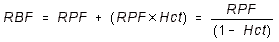
- The larger the hematocrit, the larger the renal blood flow.
- CREATININE CLEARANCE: Clinically Creatinine clearance is measured to estimate
GFR, instead of Inulin clearance.
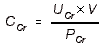
- NUMERATOR is falsely raised a little because some secretion of creatinine
occurs in the kidney.
- DENOMINATOR is falsely raised a little because of non-creatinine chromogens
the react with the creatinine testing reagent, in the blood.
- The two offset each other, so Creatinine clearance is generally considered to be
a good indicator of GFR.
- Falsely high GFR values may be obtained with people who have good blood flow
(RPF) but poor glomerular function (GFR).
- FRACTIONAL EXCRETION: The fraction of the filtered amount of a substance that
the tubule excrete. This is a measure of reabsorption capacity. The smaller the
fractional excretion, the better the reabsorption capacity.
Fractional Excretion of Water: 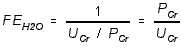
- All you have to do is measure Creatinine in the blood and in the urine and
take the ratio.
- The lower the Fractional Excretion of water, the better. A low fractional
excretion indicates that tubular reabsorption functions are working.
- FRACTIONAL EXCRETION of Any Other Substance:
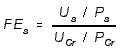
- You Pee / You Pee is the mnemonic to remember this.
- Again, higher fractional excretion indicates impaired tubular function.
- FRACTIONAL EXCRETION OF SODIUM:
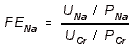
- FENa should be 1% - 3%. Anything higher than 3% indicates impaired tubular
function.
- Diuretics, of course, will falsely make this number a lot higher.
- FRACTIONAL REABSORPTION RATE = (1 - Fractional Excretion) for any
substance.
- The higher the Fractional Reabsorption, the better.
- PLASMA CREATININE CURVE: High Plasma Creatinine means low creatinine
clearance, which means trouble. Taking the reciprocal (1 / PCr) of PCr will tell you how
a chronic patient is improving.
- If the reciprocal is decreasing rapidly over time, then the patients condition is
worsening.
- If the reciprocal is leveling off in its decrease, then the patient is slowly improving.
Return to top
REGULATION of RENAL FUNCTION
UREA: It is freely filtered, and its reabsorption is dependent on urine flow rate.
- The higher the urine flow rate, the less of it is reabsorbed.
- Permeability to Urea occurs in two places:
- PROXIMAL TUBULE: Some urea reabsorption occurs, but more water reabsorption occurs so urea filtrate concentration actually goes up.
- INNER MEDULLARY COLLECTING TUBULE: Due to concentration prior to this
point, a large gradient for Urea reabsorption occurs in this segment. Urea is
reabsorbed and concentrated into the interstitial medulla, where it plays an integral
role in counter-current exchange.
- Uremia: Renal failure makes urea accumulate in the blood. However, urea is not as
toxic as some other metabolites that accumulate, so uremia toxicity usually isn't due
to urea per se.
DIURESIS: An increase in water excretion.
- Water Diuresis: Increased water excretion without corresponding increase in salt
excretion.
- Primary cause = increased intake of water.
- Increased water intake will cause plasma ADH levels to fall.
- Diabetes Insipidus = water diuresis resulting from no ADH secretion (usually)
or faulty ADH receptors.
- Water diuresis only exerts its effects on the distal tubules. That's where ADH can
exert influence.
- Thus water diuresis fractional excretion never exceeds 8% - 11% of GFR.
- Osmotic (Solute) Diuresis: Increased water excretion concurrent with increased salt
excretion.
- Causes:
- Massive increase in salt present in the tubular fluid.
- Diuretic drugs ------> inhibited reabsorption
- Solute diuresis can act at any specific site where reabsorption is impaired or
inhibited.
REGULATION OF PLASMA OSMOLARITY:
- ADH-SECRETION: Primary mechanism that respond to plasma osmolarity.
- Produced in Supraoptic and Paraventricular Nuclei of Hypothalamus ------>
Posterior Pituitary
- FEEDBACK MECHANISM:
- STIMULUS: Rise in ECF Osmolarity ------> ADH Secretion from
posterior pituitary.
- Osmoreceptors in the hypothalamus sense an increase in plasma
osmolarity.
- The range over which the receptors operate is a very strict, sensitive
range.
- BASAL ACTIVITY: 280 mOsm / kg ------> 0.5 pg / mL ADH.
- MAXIMAL SECRETION: 295 mOsm / kg ------> 4.0 pg / mL ADH
- Effective stimuli for ADH release are NaCl and Mannitol -- NaCl and
water loss are the most important stimuli.
- Urea is not an effective stimulus.
- Blood glucose is not an effective stimulus when insulin is present.
In the absence of insulin there is a small effect.
- FEEDBACK: Plasma Osmolarity ------> ADH ------> Water
Reabsorption ------> Plasma Volume ------> Plasma Osmolarity
- DIURESIS: 70 kg person ingests 1L of water ------> 28% (280 mL goes to plasma)
------> very small decrease in plasma osmolarity.
- This effect is not enough to alter GFR.
- This effect is enough to reduce ADH secretion in posterior pituitary.
- ADH DISORDERS:
- DIABETES INSIPIDUS:
- Primary Insufficiency of ADH is the most common cause.
- Nephrogenic Diabetes Insipidus is inability for kidney to respond to ADH.
- Psychogenic Diabetes Insipidus is compulsive water-drinking (polydipsia).
- SIADH: Syndrome of Inappropriate Secretion of ADH. Excessive ADH secretion.
REGULATION OF PLASMA VOLUME:
- ATRIAL RECEPTORS: Stretch receptors in the Right and Left Atria that respond to
high plasma volume.
- STIMULATE Atrial Receptors ------> Fire Vagus Nerve ------> Multiple end-effects.
- Suppression of ADH release in hypothalamus.
- Decreased Sympathetic Outflow ------> arteriolar vasodilation ------>
higher capillary hydrostatic pressure ------> edema (fluid moves out of
vascular space and into interstitium).
- Decreased Sympathetic Outflow ------> less Renin from kidney.
- ATRIAL NATRIURETIC FACTOR (ANF) is also released when the stretch
receptors are stimulated (but not via Vagus). ANF generally works to reduce
blood volume.
- ANF will cause further arteriolar vasodilation ------> edema.
- ANF inhibits Aldosterone in the Adrenal Cortex.
- BARORECEPTORS: They will decrease sympathetic outflow ------> less Renin.
- They can also inhibit ADH secretion.
- JUXTAGLOMERULAR APPARATUS:
- TWO STIMULI (INPUTS):
- Arterial Pressure Changes (Afferent Arteriole)
- Rate of flow of tubular fluid (i.e. rate of delivery of salt) in Macula Densa of
the DCT.
- MULTIPLE RESPONSES (OUTPUTS)
- Sympathetics can influence the JGA by resetting the set-point of tubulo-glomerular feedback ------> Alter arteriolar constriction and perhaps
mesangial cell constriction.\
- SYMPATHETICS: An increase in sympathetics will result in more water retention, via
peritubular capillaries, in the kidney and will result in a higher filtration fraction.
- Sympathetics ------> ------> GFR / RBF ratio ------> capillary
oncotic pressure ------> more water reabsorption.
- This is a direct JGA effect, as well as via Renin and Angiotensin (see
below).
- Sympathetics ------> ------> GFR / RBF ratio ------> Capillary oncotic
pressure ------> more water excretion.
- alpha-RECEPTORS:
- Vasoconstriction in the efferent arteriole.
- alpha-receptors are on the proximal tubules to promote Na+ reabsorption.
- beta-RECEPTORS: beta-Receptors are on granule cells to promote renin release.
- RENIN / ANGIOTENSIN SYSTEM: Renin ------> Angiotensin I ------> Angiotensin
II
- Three pathways for controlling Renin Secretion:
- INTRARENAL BARORECEPTOR: In the afferent arteriole, a fall in pressure
------> direct stimulation of renin release in granule cells.
- SYMPATHETIC INPUT: Neural (NorE) and Humoral (Epi) input to the JGA
stimulates rein release.
- beta-Receptors on Granule cells respond to both NorE and Epi to
release Renin.
- Atrial Stretch Receptors are believed to be the major sensors involved
in this pathway -- not the baroreceptors.
- MACULA DENSA FEEDBACK:
- Reduced tubular fluid flow (reduced delivery of salt to macula densa)
------> stimulate release of Renin.
- ANGIOTENSIN II: Potent vasoconstrictor
- It stimulates release of Aldosterone.
- It stimulates NaHCO3 reabsorption in the proximal tubule ------> more water
reabsorption.
- It preferentially constricts the efferent arteriole ------> capillary pressure
and RPF ------> GFR / RPF ratio (i.e. RPF decreases more than
GFR).
- The result of this is that the peritubular capillary oncotic pressure, PIb,
increases ------> more water reabsorption in proximal tubule.
- ALDOSTERONE:
- Its release is stimulated by Angiotensin II and K+ in blood.
- It promotes Na+ reabsorption and K+ excretion in distal tubule.
- ADH: ADH also responds to low blood volume (blood pressure) directly. This
response is much less sensitive than the response to osmolarity.
- ATRIAL NATRIURETIC FACTOR: It reduces blood volume by increasing the excretion
of salt and water (mechanism unknown).
- It is known to supress Aldosterone secretion, and it may supress renin secretion
too, but the evidence is conflicting.
SEVERE VOLUME DEPLETION: Summary of volume effects
- MAJOR STIMULI:
- Increased plasma osmolarity
- Decreased blood volume (atrial receptors)
- Decreased blood pressure (baroreceptors)
- MAJOR OUTPUT
- SYMPATHETICS
- Increase in Renin / Angiotensin / Aldosterone system
- KIDNEY: Reduced excretion of salt and water; increased retention.
ACUTE VOLUME EXPANSION: Summary of volume effects
- MAJOR STIMULI: Change in atrial volume (volume receptors) and atrial pressure
(baroreceptors)
- MAJOR OUTPUTS:
- Atrial Natruretic Factor is probably the most significant positive response.
- Decreased Sympathetics
- KIDNEY: Massive diuresis and dilute urine
ACID-BASE BALANCE: Usually, net secretion of acid occurs (we have an acidic diet).
- PROXIMAL TUBULE: HCO3- is the predominant buffering system acting.
- Na+/H+ ANTIPORT is stimulated by an acidic (less than 7.4) pH ------> secrete
more acid.
- It is inhibited by a basic (greater than 7.4) pH ------> secrete less acid.
- HCO3- REABSORPTION occurs in the proximal tubule to the greatest extent
(about 75%)
- However, because it is a low gradient system, it still does not affect the tubular
fluid pH that much.
- DISTAL TUBULE: NH3 and HPO4-2 are the predominant buffering systems.
- ACID PUMP: H+-ATPase secretes protons into the tubular fluid.
- These pumps are located in alpha-intercalated cells in the distal tubule.
- Transporters that balance the H+ pump:
- Cl-/HCO3- ANTIPORT gets rid of the excess HCO3- created by this
proton pump. It pumps the HCO3- out into the blood and Cl- into the cell.
- Cl--Channel, finally, then recycles the Cl- back out into the blood as well.
- REGULATION OF BLOOD pH: Blood PCO2 ------> H+ concentration
in cytoplasm of tubular cells ------> H+ secretion.
- ELECTROCHEMICAL GRADIENT: The H+ pump is limited by the electrochemical gradient.
- H+ in tubular fluid ------> hyperpolarize apical membrane ------> turn
off pump.
- The limit of H+-excretion by this pump is reached at a tubular pH of
about 4.5
- BASE PUMP: Cl--HCO3- ANTIPORT pumps HCO3- into the tubular fluid.
- beta-Intercalated Cells are the names of the cells that secrete base.
- This is the exact reverse of above. The H+-ATPase pump is then put on the
basolateral membrane to counteract the HCO3- pump.
- OVERALL CAPACITY: The distal tubule can secrete very acidic urine. However,
the presence of any HCO3- in this segment will lessen its ability to secrete acid
as the non-bicarbonate buffers become weaker.
- TUBULAR FLUID BUFFERS: An increase in the amount of buffers in the tubular fluid
will increase the rate of H+-Secretion. All secreted protons must be buffered in the
tubular fluid.
- BICARBONATE H2CO3 : HCO3- BUFFER: Base reabsorbed in the form of HCO3-.
This mechanism predominates in the proximal tubule.
- BUFFERING PROCESS:
- H+ reacts with HCO3- in tubular fluid ------> CO2 + H2O
- CO2 then comes back into the cell.
- Inside the cell, the CO2 reacts with H2O and Carbonic Anhydrase to
reform HCO3-
- HCO3- is then excreted into blood.
- NET RESULT: Movement of one HCO3- from the tubular fluid to the
blood ------> net excretion of acid.
- PLASMA HCO3- CONCENTRATION determines the filtrate HCO3- concentration, which determines how much buffering capacity the filtrate will have.
- FEEDBACK: Plasma [HCO3-] ------> Filtrate [HCO3-] ------>
H+-secretion and higher blood pH.
- Plasma [HCO3-] below 24 mEq/L: Almost all HCO3- will be reabsorbed
to keep HCO3- levels in blood higher.
- Plasma [HCO3-] above 24 mEq/L: Both reabsorption and excretion of
HCO3- increase, but excretion increases even more ------> net
excretion of HCO3-
- PHOSPHATE HPO4-2 : H2PO4- BUFFER: Acid excreted in the form of H2PO4+.
This mechanism predominates in the distal tubules.
- When HPO4-2 picks up an H+, it is still negatively charged afterward, which
means it remains lipid-insoluble and it must be excreted.
- This is a non-bicarbonate buffer and is responsible for acid secretion. The
rate of acid-secretion will be proportional to the strength of this buffer.
- AMMONIA NH3 : NH4+ BUFFER: Acid excreted in the form of NH4+. This
mechanism predominates in the distal tubules.
- As above, once NH3 accepts a proton to become NH4+, it is charged and lipid-insoluble, and thus it must be excreted.
- This is a non-bicarbonate buffer and is responsible for acid secretion. The
rate of acid-secretion will be proportional to the strength of this buffer.
- KIDNEY PRODUCTION of AMMONIA: GLUTAMINASE provides the kidney
with the majority of ammonia that it excretes. It will yield ammonia +
Glutamate, from Glutamine. Further deamidation of glutamate (to alpha-ketoglutarate) also provides some ammonia.
- Both these rxns occurs in tubular cell mitochondria.
- NH4+ can also be actively transported in place of H+ in the Na/H antiport, and
in place of K+ in the K/Cl/Na loop transporter.
- ACID-EXCRETION: NON-BICARBONATE BUFFER CONCENTRATION ultimately
determines the ability of the kidney to excrete acid!
- Plasma HCO3- Concentration: High plasma HCO3- ------> High filtrate HCO3-
concentration ------> the HCO3- buffer becomes relatively stronger than the NH3
and HPO4- buffers in the tubular fluid ------> less acid excretion.
- Blood PCO2: High blood PCO2 ------> ------> greater acid excretion by two
different mechanisms.
- SUMMARY: Low blood HCO3- and high blood PCO2 both lead to greater acid
excretion.
- CARBONIC ANHYDRASE: It maintains a constant pH within tubular cells.
- ACETAZOLAMIDE inhibits Carbonic Anhydrase ------> less H+ secretion in
proximal tubule (inhibit Na+/H+ antiport) and less Na+ reabsorption ------>
diuresis.
- Acetazolamide is thus a diuretic that acts by inhibiting carbonic anhydrase.
- Continued use will cause secretion of an alkaline urine and will result in
acidosis.
COMPENSATED RESPIRATORY ACIDOSIS: Example of kidney acid-base regulation.
- HYPOVENTILATION ------> PCO2 ------> [HCO3-] ------> pH
- The higher PCO2 in tubular cells stimulates the secretion of acid. This will correct for
the acidic pH but not the plasma HCO3- concentration.
- OVERALL: Chronic Hypoventilation, as in COPD.
- pH near normal 7.35
- HCO3- extremely high 34-36 mEq/L
- PCO2 high (hypercapnia) 60 mm Hg
METABOLIC ALKALOSIS: As from chronic vomiting.
- RESPIRATORY RESPONSE is chronic hypoventilation ------> pH is completely
corrected but a higher HCO3- results.
- KIDNEY RESPONSE: plasma [HCO3-] ------> HCO3- in filtrate ------> both
excretion and reabsorption of HCO3- increase, but excretion increases more ------>
net alkaline urine is created.
POTASSIUM HOMEOSTASIS: The kidney is the main organ responsible for potassium
balance.
- EXTRARENAL MECHANISMS:
- Insulin stimulates Na/K-ATPase in liver and muscle ------> K+ uptake
- This is a mode of absorbing dietary potassium, in the absorptive state.
- Aldosterone acts on extrarenal tissues to increase K+ cellular uptake, but this
is a slow mechanism.
- Catecholamines (beta2) indirectly stimulate Na/K-ATPase
- Acidosis ------> blood K+ by shifting K+ to the ECF because of the shift in
electrochemical gradient.
- KIDNEY
- REABSORPTION: Over 90% of filtered K+ is reabsorbed.
- PROXIMAL TUBULE (65-70%): Mechanisms are not completely understood
here; much probably occurs via paracellular diffusion.
- LOOP of HENLE (20%): Na/K/Cl transporter.
- SECRETION: Basically all K+ in the urine came from secretion in the distal and
collecting tubules.
- PRINCIPLE CELLS in the DCT and COLLECTING TUBULES: They secrete
K+.
- Factors affecting K+ Secretion:
- Plasma K+ concentration directly drives the Na/K-ATPase ------> more K+ into
Principle cells ------> more K+ secretion.
- ALDOSTERONE promotes K+ secretion in the principle cells.
- It is thought to do this by stimulating production of the Na/K-ATPase pumps.
- Again, this is a long term-effect.
- ADH raises K+ permeability to stimulate secretion. Effect not as significant as
Aldosterone.
- Alkalosis increases K+ membrane conductance in apical channels by increasing
the pH of the tubular fluid.
- DIURETICS have important effects on K+ secretion. They potentially lead to
hypokalemia which is an unwanted side-effect of diuretics.
- Carbonic-Anhydrase Inhibitors are potent stimulators of K+-secretion.
- Thiazide diuretics increase tubular fluid flow rate and thus K+ secretion.
- Loop diuretics increase K+ excretion because they inhibit K+ reabsorption.
Return to top