 Return to Biochemistry
BIOCHEMISTRY -- GENETICS AND MOLECULAR BIOLOGY
Download a copy of this study guide
 Return to top
BASIC DNA STRUCTURE
Mice Experiments:
- DNA TRANSFORMATION
- Three groups:
- Type R (rough non-virulent)
- Type S (Smooth virulent), but heat-killed.
- A mixture of both type R and type S
- Results: In group 3 the mice died because the other bacteria took up the intact DNA
-- transformation.
- TRANSGENIC EXPERIMENTS:
- Inject a plasmid containing the gene for growth hormone into a mouse-egg nucleus.
- The host-genome can take up the plasmid and express growth hormone, resulting in
big mice.
Basic DNA Structure:
- The purines have two rings. The pyrimidines have one ring.
- Purines: 6C-Ring + 5C-Ring
- Adenine has an NH2 on the six-membered ring
- Guanine has an NH2 and carbonyl on the six-membered ring.
- Pyrimidines: One 6-membered ring with two nitrogens on it.
- Thymine has two carbonyls with a nitrogen in the middle.
- Cytosine has a double bond with nitrogens flanking both sides.
- Nucleoside: The nitrogenous base + the deoxyribose sugar.
- Nucleotide: Nitrogenous base + the sugar + phosphate group.
- Nucleotide names: adenosine, thymidine, cytidine, guanosine.
- Sugar Phosphate Backbone: 3' 5' are positions on the ribose sugar.
- Phosphate groups hook on at the 5' carbon -- i.e. the 5th carbon of the ribose ring
(where the ribose is hooked to the base at the first carbon).
- Phosphates hook to each other by hooking the 5' phosphate of one sugar to the 3' OH
of the next sugar.
- Deoxyribose refers to missing OH group at the 2'.
- Base-pairing:
- A-T forms two hydrogen bonds.
- C-G forms three hydrogen bonds.
- Double-helix:
- DNA-strands are antiparallel. The template strand goes 5' ------> 3', while the
anti-template strand goes 3' ------> 5'.
- There are 3.4 angstroms between each base-pair, and 10 base-pairs in one turn of the
double-helix.
- Stability of the Double-Helix
- Hydrophobic Interactions: The ribose-phosphate is hydrophilic on the outside of the
chain. The bases are hydrophobic on the inside.
- Stacking Interactions: Van der Waals forces. Relatively weak but additive. Caused
by the planar nature of the bases.
- Hydrogen Bonding: Actually not the strongest of interactions. Facilitates stacking.
- Electrostatic Interactions: Both within chains and between chains. Phosphate groups
try to get as far away from each other as possible.
- Length of DNA:
- All DNA in human genome would cover one yard.
- About 1 x 10⁸ base pairs in one metaphase chromosome.
DENATURATION OF DNA: Separating the DNA strands
- Denaturation rate (i.e. the amount of denaturant required to do the job) depends on adenine
solubility -- not on the relative number of double and triple hydrogen bonds.
- The more soluble adenine is in the denaturant, the less reagent required to denature.
- AT-Rich region melt first because they have only two bonds.
- Heat, pH, and temperature extremes will also destabilize and thereby denature DNA.
- Hyperchromicity: The way to detect denatured DNA. It absorbs stronger in the 260 nm
region due to presence of more nucleotides.
- Tm, Melting Temperature: The temperature where half of the DNA is denatured. This
value is characteristic of the relative amount of A-T and G-C present.
- The more G-C present, the higher the Tm (i.e. the higher the temp required to
denature).
- DNA denatures from many points at once. When you look at an EM and see a bubble of
single stranded, you know it's A-T rich because that's the part that denatures first.
RENATURATION OF DNA: To renature the DNA, bring the temperature back down.
- Two general steps of Renaturation:
- Nucleation: The random association and dissociation of strands. This is the slow
step.
- Zippering: After nucleation has occurred, the strands are oriented and the rest of the
renaturation can be completed relatively quickly.
Rate-Constant Equation: 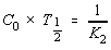
- C0 = Sequence Concentration.
- T1/2 = Time for half-reaction
- K2 = 2nd order rate constant.
- COT Curve (Renaturation Curve):
- Not ideal 2nd order kinetics. The curve is bumpy.
- Initial foldback phase = renaturation where repetitive sequences anneal with
themselves.
- Intermediate Phase:
- Single-Copy Phase: Single-copy genes re-anneal to each other.
- Sequence Concentration: The relative amounts of the same sequences present in a given
amount of DNA. Repetitive sequences yields high sequence concentration.
- E-Coli have about 1000x higher sequence concentration than humans.
- Sequence concentration is directly proportional to rate of renaturation. The faster
the renaturation, the higher the sequence concentration.
- If given 1 mg each of E. Coli and Human DNA, the E. COLI would have 1000x more
copies of each gene present, because its overall genome-size is smaller.
Different Types of DNA:
- Single-Copy DNA (75% of genome): Gene-sequences found basically in only one place in
the genome.
- Repetitive Dispersed DNA Fractions (15% of genome): Characteristic repetitive sequences
found at disparate places throughout the genome.
- Alu sequences: A family of repetitive sequences.
- Around 300 bp long.
- Around 300,000 copies of the gene at disparate places in genome.
- Satellite DNA (10% of genome): Highly repetitive sequences localized to the centromere and
telomere, usually.
HISTONES:
- There are five histones: H1, H2a, H2b, H3, H4
- Histones are basic -- rich in lysine and arginine
- Every bead in DNA has eight histones -- two tetrameric molecules, each consisting four
histone-subunits.
- DNA turns 1 3/4 times around each bead, to form the nucleosome core.
- H1 is on the outside and finishes off the 1 and 3/4 turns, to make it 2 full turns per
chromatosome.
DNA PACKAGING: Histones aid in DNA packaging
- Chromosomes are least dense during interphase, when transcription is taking place.
- Chromosomes are most condensed during metaphase, when they are packaged tightly with
histones.
DNA SEQUENCING: Dideoxy chain termination protocol
- Set up four tubes, one of each dideoxy nucleotide.
- Each of the four tubes contains DNA wanting to be sequenced, DNA-Polymerase,
and one of each dideoxy nucleotide.
- You must start with a primer to initiate DNA synthesis.
- The dideoxy nucleoside has no hydroxy in the 3' position, so that it cannot form
phosphodiester linkages. As a result, DNA synthesis may cut short at each sequence for the
specified tube.
- So for ddATP, at every adenine, the polymerase might add (1) Adenine triphosphate,
or (2) the Dideoxy-Adenine triphosphate.
- RESULT = all possible chains of different lengths, with the specified nucleotide ending at
each chain.
- VISUALIZATION: You can then line up A, T, C, and on a PAGE gel, and visualize each
respective chain length in order, distinguishing exactly one nucleotide for each segment-length.
- To read the gel, start at the bottom -- shortest fragment -- and go to each next longer
fragment. You can literally just read off the nucleotide sequence according to
fragment length.
- AZT: AZT is a chain terminator, like the dideoxy nucleotides.
- It is not phosphorylated so it can get into cells. It is phosphorylated once into cells.
- It is an analog of thymidine. Reverse Transcriptase will incorporate it into a DNA
chain, but once incorporated, reverse transcription stops because it can't form another
phosphodiester bond.
- There is some toxicity involved since some of it may be incorporated into the cellular
machinery.
GENE STRUCTURE: Exons / Introns
- Exon: The nucleotides of a gene that are actually translated.
- Some genes can have many disparate exons composing one gene.
- Factor VIII, e.g., is a very large gene -- it has 26 exons.
- Exons are very small in nucleotide length compared to the intervening introns.
- Introns: Intervening nucleotide sequences that are transcribed, but are spliced out before
translation.
- Familial Hypercholesterolemia: Autosomal dominant familial high cholesterol.
- Normal cholesterol = 200 mg/dl
- Heterozygotes for the LDL-Receptor Defect = ~400 mg/dl
- Homozygous Hypercholesterolemia = ~800 mg/dl
- The LDL-Receptor Gene has Alu Repeats in the introns. The genetic error results from
unequal crossing over, due to those Alu Repeats.
- Occurs due to incorrect lining up of the Alu Repeats during homologous
recombination.
- One homolog may have a different number of Alu Repeats in its introns than the other
homolog. But due to sequence similarity, the two homologs may line up and
recombine according to the repeats rather than to the expressed gene.
- For LDL-Receptor, the unequal crossing over makes you lose Exon 5.
Return to top
DNA REPLICATION AND REPAIR
DNA REPLICATION:
- Two rules to commit to memory.
- DNA Polymerase requires a primer with a free 3' OH end.
- Synthesis always occurs in the 5' ------> 3' direction.
- Discontinuous Replication:
- There are multiple replication origins.
- There is one continuous leading strand, which synthesizes DNA continuously in the
5' ------> 3' direction.
- There is one discontinuous lagging strand, which synthesizes DNA in fragments,
from the 5' ------> 3' direction, and then translocates backwards, and then continues
synthesizing.
- Initiation: in E. COLI
- DNA-A binds to the origin of replication (Ori-C) and locally melts (denatures) DNA.
Initial DNA-A binding allows for more DNA-A binding until a significant amount of
DNA is complexed and unwound.
- Primosome: Primase bound together with DNA-B and DNA-C
- DNA-A allows for binding of DNA-B and DNA-C to the DNA, which in turn
starts replication.
- Primase (DNA-G) is an RNA-Polymerase which synthesizes an RNA primer
to start DNA-Replication.
- The primosome activity requires ATP (to prime the reaction).
- The lagging strand must remove the original primer in order to complete synthesis,
so there are many more steps to lagging strand synthesis than to continuous synthesis.
- Synthesis continues as it makes a replication fork.
- Okazaki Fragments: The name of the short discontinuous fragments made by the lagging
strand during DNA synthesis in the anti-parallel direction.
- Prokaryotic Replication Proteins:
- DNA-A,B,C required for initiation at the origin.
- DNA-G (Primase) synthesizes the RNA primer.
- DNA-Polymerase I -- removes the primer and fills in the gaps.
- It uses a 5' ------> 3' exonuclease activity to nibble away to the primer.
Then it fills in the gap behind itself with the needed nucleotides.
- DNA-Ligase: Fills in the last gap by forming the last needed phosphodiester bond.
Eukaryotic DNA-Polymerases:
- DNA-Polymerase-alpha -- Carries out lagging strand synthesis.
- DNA-Polymerase-delta -- Carries out leading strand synthesis.
- DNA-Polymerase-beta -- Carries out DNA repair.
DNA Mutations:
- Types of Mutations:
- Genome Mutations: Aneuploidy
- Chromosome Mutations: Translocations, incorrect recombination.
- Gene Mutation:
- Deletion
- Addition
- Point mutation
- Nonsense mutation
- Mutation Rate: The larger the gene, the more likely it is that something can go wrong with
it, the higher the mutation rate for new mutations.
- Polycystic Kidney Disease has a very high mutation rate for new mutations.
- Little sperm carry lots of mutations. 1:10 carries a new deleterious mutation.
Fortunately most of these are do deleterious as to be lethal.
- DNA REPAIR:
- It is what keeps mutations as low as they are.
- There is a correlation between DNA-Repair capabilities and lifespan. we have a
higher DNA-Repair rate than rats, so we live longer than them.
Mechanisms for Mutation to Occur
- Tautomeric Forms of DNA Bases: One way that DNA mutation (mis-pairing) can occur.
- A and C (purines) can incorrectly tautomerize from the amino form to the
imino form, changing each one's base-pairing behavior.
- G and T can incorrectly tautomerize from the keto form to the enol form, changing
each one's base-pairing behavior.
- MUTAGENIC REACTIONS
- Deamination: Oxidative changing of chemical structure, all of which change base-pairing behavior.
- Cytosine ------> Uracil
- Adenine ------> Hypoxanthine
- Guanine ------> Xanthine
- They can react with hydroxylamine. (NH2OH)
- Alkylation can occur via DMS (dimethyl sulfate)
- Perpetuation of Mutation: One round of DNA replication can make a mutation permanent,
by correctly base-pairing it the next time around, and then it is no longer subject to repair and
it's stuck that way.
- Oxygen Radicals: Radicals can knock out genes. Anti-oxidants can fight this. Iron oxide can
react with DNA and cleave it.
- Aflatoxins are oxidative substances that give rise to cancer. Anthracene is
carcinogenic substance found in cigarette smoke.
- UV-Light causes thymines to dimerize on a strand where two thymines occur next to each
other, forming a cyclobutane ring on only one of the strands, ruining replication.
- Intercalating Agents are agents that squeeze in between the bases, causing deletions and
insertions.
- Ethidium Bromide is an intercalating agent.
DNA REPAIR MECHANISMS
- Excision Repair: Damaged base causes excision enzymes to remove that base so it can be
put on again correctly.
- DNA-Poly-I performs this repair mechanism, then ligase seals the nick.
- Depurination Repair: Depurination means you lose a (purine) base -- i.e. a deletion, except
that the backbone remains and only the base is gone.
- Repair = an endonuclease will cut out the empty backbone, and DNA-Poly-I +
Ligase can refill and seal the nick.
- DNA Glycosylase is a special enzyme that recognizes and removes Uracils whenever they
pop up in DNA. Then repair takes over as usual.
- Uracil winds up in DNA usually due to the spontaneous deamination of cytosine.
- Deamination of 5-methyl cytosine cannot be repaired! 5-methylcytosine can be found at
regions of high gene activity (near promoters).
- Deamination of 5-methyl cytosine results in Thymine, which is not foreign, so it isn't
repaired.
Return to top
RNA and TRANSCRIPTION
Modified Bases: There are many modified bases in RNA, especially in tRNA. Nuff said.
RNA Structure:
- Mostly single stranded.
- The sugar is ribose. It has a 2' OH group. This makes the phosphodiester bond more labile,
which makes RNA more unstable than DNA. So it doesn't last as long!
- mRNA Structure:
- A 7'-Methyl GTP CAP at the 5' end.
- The 5' Untranslated Region.
- The Initiation Codon (methionine)
- The coding region
- The 3' Untranslated Region.
- The end of the untranslated region has a polyadenylation signal, which tells the
RNA Polymerase to add a Poly-A Tail.
- mRNA Populations:
- The more specialized cells (e.g. mouse liver) have very few different mRNA's in huge
quantity.
- The less specialized mrna's -- housekeeping cells have a vast number of different
mrna's, each one transcribed only a few times.
- Heterogenous Nuclear RNA, hnRNA = the RNA right after it is transcribed -- before the
introns are removed, and before any other post-transcriptional modifications are made.
- Basic tRNA Structure: It has a secondary structure -- the local folding domains, containing
the anti-codon and amino-acyl binding sites.
- It also has a tertiary structure, which actually interacts with the ribosome.
TRANSCRIPTION
- Initiation of Transcription: RNA-Polymerase binds at the promotor, changing the closed
promotor complex into an open promotor complex.
- E. COLI -- RNA-Polymerase recognizes two consensus sequences upstream from the start-site of replication. RNA polymerase binds both of the following at the same time. It scans
down the DNA looking for these sequences.
- TATA-Box, about -5 base pairs from replication.
- TCTTGAGAT-Box, about -35 bp from replication origin.
- EUKARYOTES -- Promotor Regions
- Also have a TATA-Box often. Its function is to control the starting place for
transcription.
- If you remove TATA from the gene, the transcription rate does not go down,
but the starting point becomes sloppy. So it directs the polymerase where to
start transcribing.
- Often find GC-Boxes -- GC-rich regions.
- Types of Eukaryotic RNA-Polymerases
- RNA-Polymerase I -- Ribosomal RNA (rRNA)
- Insensitive to alpha-amanitin.
- RNA-Polymerase II -- Messenger RNA (mRNA)
- Extremely sensitive to alpha-amanitin.
- RNA-Polymerase III -- tRNA and 5s rRNA
- Sensitive to alpha-amanitin, but only at high levels.
- alpha-Amanitin is a transcription inhibitor that can be used to distinguish the different
RNA's.
- Yeast RNA Poly II has multiple subunits, some of which are shared in common with other
polymerases. One of the ones in c
SIGMA FACTOR -- it allows the polymerase to scan the DNA more efficiently and quickly to find
the promotor.
- It decreases the polymerase's affinity for DNA so it can scan faster.
- At the same time, it increases polymerase's affinity for the promotor region itself.
- Sigma factor is specific to prokaryotes.
PROKARYOTIC INITIATION OF TRANSCRIPTION:
- RNA-Polymerase II binds to the promotor region. Sigma factor is intact. It transcribes off
of the anti-template strand in a 5' ------> 3' direction.
- It puts the first nucleotide on. It is trial and error which nucleotide comes on, but when the
right one does, it stops and sticks.
- First phosphodiester bond is formed.
- Sigma factor is released!
- RNA-Polymerase translocates to next base.
EUKARYOTIC INITIATION OF TRANSCRIPTION: Realize that there are lots of others
transcription factors (such as jun and fos) that act in conjunction with RNA-Polymerase, in
eukaryotes.
Factor IX Gene Promotor: The promotor for factor IX can get mutated, leading to a lower level
of gene expression of Factor IX (but not a malfunctional gene itself) ------> leads to hemophilia.
Return to top
MRNA PROCESSING / SPLICING
Order of events after our hnRNA is made:
- 7'-methyl-GTP CAP is put on. The guanosine is methylated.
- Poly-A Tail is added next, about +30 from the adenylation signal -- AAUAAA.
- The mRNA Polymerase is transcribing, and when it reaches this signal, it cleaves the
transcript at that point.
- Then, the Poly-A-Polymerase takes over and puts on lots of adenines as a tail.
- Then mRNA splicing occurs.
Chemistry of mRNA Splicing:
- The free 2' OH of an internal adenosine residue within the intron attacks a terminal
guanosine, breaks the phosphodiester bond, and hooks onto it.
- So the intron loops with itself, hooking the internal A to the terminal G.
- The intron product forms a lariat structure -- a loop with a tail on the end of it.
- This leaves a free 3' OH on the terminal Guanosine of the exon, which then attacks another
guanosine, in same fashion, at the other end of the intron -- which links it to exon 2.
Spliceosome: The protein assembly that accompanies mRNA splicing.
- Specific sequences always border the intron -- a GU on the 5' side and an AG on the 3' side,
and there is always an A in the middle.
- Small Nuclear Ribonucleoproteins (snRNP's) bind to the RNA to do the splicing.
- Structure = protein subunits U1-U6 + small segments of RNA.
- FUNCTION: They help to recognize the starting and ending points of splicing. U1
binds to 3' end and U5 binds to 5' end. They also help to stabilize the lariat structure.
U2 recognizes the internal adenosine.
- Once splicing is complete, the snRNP's are sloughed off along with the lariat-intron
structure.
- Consensus structures at the splicing site:
- 100% of discovered cases have GT (GU) at the begging of the intron and AG ( at the
end). That is a 100% consensus sequence.
- Commonly there is a polypyrimidine (TC) track in the middle -- high frequency of that
in discovered introns.
- There are other consensus sequences (of varying recurrence frequency) throughout,
which the spliceosome probably uses to identify where to splice.
Beta-THALASSEMIA: Thalassemia simple means underproduction of the beta-globin gene. The
beta-chain gene contains three exons and two introns. Clinically it results in hemolytic anemia.
- beta+-Thalassemia: A decrease in beta-Chain Production. Two different examples. In both
beta+ cases, the original splice site remains unmutated.
- One example: A new splice site is created within the intron by a point mutation. The
spliceosome cuts at that site 90% of the time, yielding only 10% functional beta-chain.
- Second example: A point mutation that activates a new splice site by mutating the
base +1 from the GT invariable sequence. It changes it to a popular consensus base
for that position, making the spliceosome recognize it when it didn't before.
- beta0-Thalassemia: No beta-Chain production. beta0 case -- the original splice site is
mutated.
- This can be caused by a splicing error, where the normal splice site for one of the
beta-chain exons is mutated, forcing the spliceosome to cut at a cryptic site (formerly
ignored) inside the intron. The result is that there is an extra piece of inton in the
expressed gene, rendering the protein non-functional.
Alternative Splicing: You can introduce variety into protein products by splicing at alternative sites.
There are different ways to achieve this:
- Cassette -- do or don't include the exon.
- Mutually Exclusive -- include either one exon or an alternative exon.
- Use alternative promotors.
- Use alternative adenylation signals.
- Tropomyosin gene does alternative splicing. Different tissues cut the tropomyosin gene in
different ways to get different forms (isozymes) of tropomyosin. So alternative splicing is a
way to achieve tissue specific proteins.
- Troponin-T is another example.
Return to top
TRANSLATION / PROTEIN SYNTHESIS
GTP -- Provides the energy source for protein synthesis.
ATP -- Provides the energy source for activation of the amino acid.
START CODON: AUG. It codes for methionine.
STOP CODONS: UAA, UAG, UGA
tRNA STRUCTURE:
- There is at least one specific tRNA for each amino acid.
- All tRNA's are 75 to 76 base-pairs in length.
- There is extensive interbase-pairing, and trna's have characteristic clover-leaf appearance.
- Amino-Acid Acceptor Stem: Always at the 3' end.
- The 3' free OH of the ribose attaches to the C-terminus of the specified amino acid.
- The 3' end of all amino acids terminates with the bases, CCA.
- Anticodon: In loop 2, in the lower region of the molecule. It base-pairs to the codon of the
mRNA to specify which amino acid to bind.
tRNA CHARGING: Occurs in two steps. First, the amino acid is activated (adenylated). Then the
amino acid is actually added to the tRNA.
- Both steps are carried out by the same enzyme -- Amino-Acyl tRNA Synthetase.
- Amino Acid Activation: AMP is added to the amino acid (adenylation), by cleavage of one
ATP, before the acid is added to the tRNA.
- Acylation of tRNA: The AMP is used to catalyze the addition of the acid onto the tRNA
at the acid-accepting site. The AMP is simultaneously sloughed back off.
- The bond energy of the ATP is held in the amino acid tRNA and is used in translation.
- Specificity: There is a unique tRNA-Synthetase for each tRNA. That confers high specificity
to the process, to prevent mispairing.
- Each enzyme binds to one of the unique sites on the tRNA -- in one of the other
loops.
THE GENETIC CODE: Okay?
- Frameshift mutation: Addition or deletion of a non-multiple of three base pairs.
- The aa sequence will be altered.
- The stop codon will invariably be changed therefore, so the resulting protein will also
invariably be (usually) shorter or (perhaps) longer than the original.
Hemoglobin Wayne: A frameshift mutation in alpha-globin near the very end of the protein.
Strange thing -- it eliminates the stop codon and results in a longer protein by five amino acids.
Read-Through / Reverse-Terminator Mutations: Mutations in the stop-codon itself, which results
in a long protein. These occur in alpha-globinopathies.
Ribosome Structure:
- Ribosome consists of a large subunit and a small subunit:
- Bacteria: Sedimentation values
- Small Subunit = 30s. Has a 16s rRNA molecule attached to it.
- Large Subunit = 50s. Has a 23s rRNA molecule attached to it.
- The whole ribosome = 70s
- Mammals:
- Small Subunit = 40s. Has a 16s rRNA molecule attached to it.
- Large Subunit = 60s. Has a 28RNA molecule attached to it.
- The whole ribosome = 80s
- Two active sites on the ribosomes:
- Peptidyl-Transferase Site: The P-Site
- Amino-Acyl Site: The A-Site
INITIATION:
- Initiator tRNA (Met) starts bound to the small ribosomal subunit.
- eIF2 is bound to the complex, too. (eukaryotic Initiation Factor)
- The initiation complex binds to 5' cap and scans down until it finds the AUG Start Codon.
When the Start Codon is reached, the initiation factors fall off.
- Once reached, the large ribosomal subunits hooks onto the tRNA, and the tRNA gets placed
into the P-Site.
- The next tRNA binds to the A-Site.
- A peptide bond is formed between the amino acids at the A and P sites.
PEPTIDE BOND FORMATION and ELONGATION: Energy comes from the ATP from tRNA-Charging.
- The free NH2 on the protein in the A-site attacks the carboxyl of the tRNA amino-acid in the
p-site.
- Enzyme peptidyl transferase carries out the reaction. It is made of 23s (or 28s) rRNA.
- The two amino-acid chain is then at the A-site, and there is an uncharged tRNA remaining at
the P-Site.
- Next translocation of the ribosome occurs, to move the chain to the P-Site, which sloughs
off the now uncharged tRNA. So after translocation we now have the growing chain the P-Site, and an empty A-site, ready for the next tRNA.
- the process repeats.
GTP Energy is required for:
- Binding of the tRNA to the small subunit of the ribosome.
- Translocation.
TERMINATION:
- Release Factors (proteins) recognize and bind to the stop codon. There is no tRNA that
recognizes the stop codon.
INTERFERON: A protein made in response to viral infections. It stimulates neighboring cells to
make anti-viral proteins. The last gasp for cells to survive viral attacks.
- The virus infects the host cell and triggers it to express interferon.
- Interferon goes to neighboring cells and induces them to express anti-viral protein (AVP).
- AVP will halt the attack, ultimately by two means: by degrading viral mRNA and by halting
protein synthesis in the host cell.
- 2-5A Synthase: This protein will create 2-5A (2'-5" Adenine RNA chain) upon attack by the virus.
- The 2-5A then activates an endonuclease that degrades mRNA, including the
viral mRNA.
- eIF2 Kinase is another anti-viral protein. It phosphorylates the alpha-subunit of e-Initiation Factor 2, halting protein synthesis and thus killing the virus.
Inhibitors of Protein Synthesis: Various agents inhibit protein synthesis by binding to different
translation factors. They are all quite specific to a site, substance, and to procaryotes or eucaryotes.
Thus many are useful as antibiotics, e.g. streptomycin, erythromycin.
Signal Sequence: A protein destined for the ER itself has a signal sequence at the N-Terminus.
Integral Membrane Proteins have signal sequences.
- Signal Recognition Particle (snrp) binds to the signal sequence and to the ER, depositing
the protein in the ER.
- When the snrp binds, the ribosome temporarily halts translation as the protein shoves
its way into the ER membrane.
- The snrp binds to a snrp-receptor on the ER-Membrane.
- A signal peptidase on the inside of the ER lumen clips the signal sequence.
N-Linked Oligosaccharides: Cotranslational glycosylation with a dolichol phosphate intermediate.
The function of carbohydrates being added in is for protein targeting, structure, and cellular
recognition.
- N-Linked oligosaccharides are linked through the amide NH2 of Asn residues. There is a
consensus sequence of Asn-X-Thr when glycosylation occurs.
- Unlike O-linked glycosylation, it occurs cotranslationally -- while translation is taking place.
- Dolichol Phosphate is a lipid-like intermediate that carries the sugar. It is on the cytoplasmic
side of the ER membrane.
- N-Acetylglucosaminyl transferase is the enzyme that glycosylates the Asn residues.
Dolichol Phosphate serves as the initial glyco-group acceptor, forming UDP-N-Acetylglucosamine. Several mannose and glucose sugars are added to it.
- All the sugars are then transferred to the amino acid as the acid traverses the membrane.
- After this initial glycosylation, additional modifying enzymes can be employed to create all
kinds of glyco groups off the basic themes.
- Glycosidases are enzymes that can trim off extra CHO's.
Targeting of Proteins to Lysosomes:
- Mannose-6-Phosphate is a carbohydrate which targets a (hydrolytic) enzyme specifically to
become a lysosome.
- I-Cell Disease: (Inclusion Bodies) -- disease resulting from defect in lysosomal M6P
receptor.
- As a result the hydrolytic enzymes don't get formed into lysosomes and no cellular
digestion occurs. The cell can lyse from toxicity and self-digestion.
- Death occurs by age 8.
Post-Translational Processing:
- There are two types:
- Proteolytic Action.
- Disulfide bond formation.
- Synthesis of Insulin: After translation is complete
- The signal peptide is cleaved
- A disulfide bond is formed between the A and B chains. The C-Chain (between the
two disulfide bonds) is then sloughed off.
- PreProInsulin
- "Pre" indicates presence of a signal peptide.
- "Pro" indicates post-translational steps are required to activate the protein.
- Preproopiomelanocortin: It may be posttranslationally cleaved at many different sites to
produce a variety of different tissue-specific proteins from the same precursor.
- Each pattern of post-translational cleavages yields a different "family" of proteins
(cell-type specific) from the same precursor.
Return to top
DNA CLONING AND LIBRARY CONSTRUCTION
Restriction Endonuclease: Cut DNA into specific fragments, around specific sequences.
- They originate from bacteria. They have restriction enzymes to cut out invading viral genes
by removing ("restricting") the viral DNA from their genome.
- Modification Enzymes: They also have modification enzymes so they don't cut out
their own DNA. These enzymes methylate the same restriction sites recognized by
the restriction enzymes, to prevent self-cleavage.
- The restriction sequences are usually palindromic.
- Sticky Ends: There are usually sticky ends that remain after cutting, and that can be re-annealed to each other.
- 5'-G|AATTC-3'
- 3'-CTTAA|G-5'
- Cut at the G, and the AATT sequence remains as a sticky end.
- Blunt Ends: No unpaired bases remaining at the end of the cleaved sequences.
- Frequency of Cutting: The longer the restriction site, the fewer fragments, on average, it will
create. Or you could say, the longer the restriction site, the less frequently it will run into that
site and cut it, on average. On the average it cuts at every 4n base pairs.
- 4-base Cutter: 256-bp fragments, on average.
- 5-base Cutter: 1024-bp, on average.
- 6-base Cutter: 4096-bp, on average.
- 7-base Cutter: 65536-bp, on average.
Forming Recombinant DNA Molecules: "Chimeric" DNA Fragments.
- You can put the sticky ends together of two fragments that weren't originally joined (but were
cut by the same restriction site), to create recombinant DNA fragments.
VECTORS: A packaging to carry DNA fragments of interest.
- Types of Vectors:
- Plasmids: Carry small amounts of DNA -- 5 to 10 kilobases. They contain three
different sties.
- At least one unique restriction site. Cut at any restriction site, and open the
plasmid at only that locale.
- An origin of replication, so it can replicate independent of the host
chromosome (like the F-Factor).
- An Antibiotic Resistance Gene, so it can be selected for.
- Bacteriophage lambda: Carries slightly larger amount -- up to 20 kilobases.
- Cosmids: -- up to 50 kilobases
- Yeast Artificial Chromosomes (YAC's): Carries the largest amount -- 100 - 1000
kilobases (i.e. 1 megabase)
- They have all the information needed to make them complete chromosomes
(centromeres, telomeres, origins of replication).
- Mostly commonly used for human genes.
HOW TO CLONE:
- Cut open a bacterial genome with a restriction enzyme such as EcoR1.
- Take your fragment of interest and use DNA Ligase to ligate it into the cut spot. The
fragment must have sticky ends that correspond to EcoR1 too.
- When done the plasmid will have two EcoR1 sites -- the original one, and the one at
the end of our new DNA-insert.
- Transformation: Insert the plasmid into bacterial cells. Treat the bacteria with Ca+2 to try
to get them to take up the plasmid.
- Grow the bacteria (and potentially the desired plasma).
- Then transfer to selective plates -- plates containing the resistance-antibiotic. The plasmids
contain the resistance gene, so the only bacteria that survive will be those who took up the
plasmid.
MILLIONS of DNA Sequences: We start with lots of DNA sequences -- all those created by the
restriction enzyme we used.
- Each plasmid will take up a different DNA sequence, and each bacteria will take up a different
plasmid.
- In the end, each bacterial colony, then, will contain identical copies of the same DNA plasmid.
Insertional Inactivation: Basically, select for bacteria by looking at the ones that were killed!
- You gotta have two Antibiotic resistance sites, and one of them must have a restriction site
in the middle of it.
- Take Amp and Tet, with a restriction site in the middle of Tet... PROCEDURE
- Take the plasmids and restriction cut them, and try to insert a DNA-Fragment into the
restriction site inside the Tet gene. This will interrupt and inactivate the Tet gene.
- Grow the bacteria on a plate containing only Amp. All surviving bacteria will have
taken up the plasmid.
- Transfer bacteria to a plate containing Tet. The bacteria who die will have taken up
a plasmid and the desired DNA fragment.
- Go back to the original plate and isolate those bacteria that died on the replica plate!
-- you have the bacteria with the desired ffragment!
Genomic DNA Library Construction: Make a bacteriophage library and put it into (i.e. infect)
bacteria.
- PREPARING THE PHAGE GENOME:
- Take the Bam-H1 (6-cutter) restriction site on the viral genome.
- Digest the viruses with the Bam-H1 endonuclease and eliminate the middle part
because it is not essential to viral replication.
- Only the two arms (the flanking segments) are essential to viral replication.
- PREPARING THE HUMAN GENOMIC DNA
- Partially digest the human DNA with Sau-3A (4-cutter).
- Sau-3A sticky ends are complementary to the Bam-H1 ends.
- Partial digestion generates multiple-sized overlapping fragments, which is
necessary for chromosome walking.
- PREPARE THE PHAGE VECTOR:
- Use DNA-LIGASE to insert the fragments into the middle part of the phage-genomes.
- Use a COMMERCIAL PROTEIN EXTRACT to package the genome into a
bacteriophage virus. The protein-coat is commercially available.
- INFECT THE BACTERIA to house the vector.
- The concentration of E. COLI must be much greater than that of the phages! --
so that, statistically, only one bacteriophage will infect each E. COLI.
- A wave of infection (clear spots -- plaques) can be seen on the bacteria-plate, as
phages undergo lytic cycle and infect neighboring bacteria, carrying the human DNA
fragment with them.
- YOU HAVE A LIBRARY -- each bacterial colony will contain a different human DNA
fragment.
Screening the Phage Library:
- Take a bacteria plate with lots of plaques on it. Each plaque is different. The plaques contain
raw DNA and dead (lysed) bacteria.
- Put a nitrocellulose filter on top of it. This smears the plaques onto the filter.
- Add a denaturing agent to denature (make single-stranded) the DNA.
- Hybridize the denatured DNA to a 32-P radioactive probe, containing the complement of the
DNA of interest. Wherever it probes will show up on a radiograph, and you will know the
corresponding plaque.
Including overlapping, over 800,000 clones (probes) are needed to represent the entire human
genome, assuming 20kb fragments.
- Theoretical minimum with no overlapping = 150,000
Life-Cycle of a RNA Retrovirus:
- Start with single-strand of RNA and reverse transcriptase protein.
- Infect host cell and use the reverse transcriptase + host machinery to make DNA from RNA.
- First you make a RNA:DNA strand, and then use the DNA host strand to make a fully
functional DNA:DNA Double-Strand.
- The DNA-fragment then inserts itself into host genome and behaves like your basic
virus.
- In lytic phase it makes more mRNA and reverse transcriptase, and packages new
retroviruses to infect other cells.
cDNA Library Construction: The purpose is to look at genes that are actually expressed -- because
we start with already-transcribed mRNA, and use reverse transcriptase to make DNA from it.
- cDNA libraries will be tissue-specific, because different tissues make different series of
mRNA.
- PROCESS:
- Take your mRNA with its Poly-A Tail. Use Reverse Transcriptase to make DNA
with poly-T at it's 5' end.
- Use an RNAse to partially digest the RNA-strand, leaving some RNA behind as
primers.
- Then use DNA-Polymerase I to make a second DNA strand so that we have double-stranded cDNA with poly-T tails.
- Then ligate on restriction sticky ends so that they can be inserted into vectors.
- Then insert the double-stranded cDNA into a vector (Restriction Enzyme + DNA-Ligase)
Return to top
PHYSICAL GENE MAPPING
REQUIRES A PROBE TO START WITH! A GENE PROBE IS BEST IF THERE IS ONE.
OTHERWISE WE NEED A MARKER PROBE FROM LINKAGE ANALYSIS.
Somatic Cell Hybridization: GOAL = to map an allele to a particular chromosome.
- HUMAN: Cannot survive if homozygous for ouabain.
- MOUSE: Cannot survive if homozygous for HAT (Hypoxanthine, Aminopterin, Thymidine)
- They are engineered to be HGPRT-, so that they can't survive if homozygous for
HAT.
- HGPRT is the salvage enzyme, necessary when de novo purine production is
blocked. Aminopterin blocks de novo purine production, so mice that are
HGPRT- cannot survive in the presence HAT.
- MIX THE CELLS together using inactivated Sendai virus.
- The only ones that survive will be HYBRIDS, or heterokaryons. Their nuclei will merge,
forming one nucleus after cell-division.
- Subsequently, some of the human cells will spontaneously be lost.
- ANALYZE THE HYBRID CELLS for traits. Take for example the HEXA gene (Tay-Sachs
Disease). It was found that all cells that were HEXA- (i.e. did not have the gene) were also
lacking Chromosome 15, and all cells that were HEXA+ did have chromosome 15.
- Chrom 15 was the only chromosome that had perfect correlation with the HEXA
gene.
- SOUTHERN BLOTTING was used to probe for the HEXA gene. You could
identify which samples had the HEXA gene by looking at the Southern blot.
- You can identify which samples have Chromosome 15 by standard karyography
techniques (cytogenetics / characteristic bands).
Southern Blotting: Look at a Electrophoresis Gel of DNA fragments.
- Do a restriction digest of human genomic DNA with a particular restriction enzyme.
- Purify the DNA and run a PAGE-Gel of it. You'll have lots of DNA all over the gel, and it
will be difficult to distinguish particular fragment-sizes.
- Use nitrocellulose and a radioactive probe to visualize a fragment of interest.
- The resulting nitrocellulose filter will contain only the DNA-fragments of interest.
- Results:
- You can compare the characteristic fragment-lengths to known lengths, by looking
at a restriction map.
- You can see whether the site was homozygous or heterozygous by noting whether the
probe resulted in two spots or only one spot.
Regional Mapping with Translocation: You can use translocated chromosomes to localize a gene
to particular region, by looking at the region of overlap between two translocated chromosomes.
- Take translocated chromosomes and make hybrids out of them (somatic cell hybridization).
- If the gene is present in the translocated chromosome, then it is known to reside in the portion
of the chromosome retained in the hybrids. Conversely, if it is absent from the translocated
chromosomes, then it is known to reside in the portion that is absent.
Gene Dosage Mapping:
- For X-linked chromosomes, we can look at the people who have polyploidy of the X-chromosome. If a southern blot for the desired gene then shows up as a more intense spot
for the polyploidy-X sample, then we know this gene is on the X-Chromosome.
Fluorescence In Situ Hybridization (FISH): If you have a probe that you wanna map, you can
fluorotag it, denature it, and probe it to a human genome. Then visualize it under fluorescent light
and see which chromosome it hybridizes to. It will fluoresce under the light.
- You can even use this method to localize the probe to a particular region in the chromosome,
depending on how well you can see exactly where it hybridizes.
- You probe to human metaphase chromosomes, so you can see where it hybridizes!
YAC (Yeast-Artificial-Chromosome) Clones: They contain overlapping piece of DNA, and can
carry the largest amounts of DNA -- up to 1000 kilobases.
Return to top
RECOMBINATION AND LINKAGE
Homologous Recombination: Crossing over between homologous chromosomes. The rate of
cross-over is directly proportional to the distance between the chromosomes, i.e. the further the two
genes are apart, the more likely it is that a recombination event may occur between them.
- EFFECT: Rather than getting all of one chromosome or all of the other, an offspring is more
likely to get some recombined combination of the two chromosomes.
- One more generation down, and the grandchild will have a mosaic of both the parents'
and the grandparents' chromosomes.
Allele: One of the alternative versions of a gene that may occupy the same genetic locus.
Homologous chromosomes contain (possibly) different alleles of the same gene.
Independent Segregation: Given mother A/a and Father B/b, the likelihood of receiving any
particular allele is 9:3:3:1 for each dihybrid phenotype, or 1:1:1:1 for each dihybrid genotype.
Completely Linked: Given maternal chromosome A/B and paternal chromosome a/b, all prodigy
are either A/B or a/b, indicating that no recombination is occurring between the A and B alleles.
Linked: The rate of recombination between the alleles is proportional to the distance between the
loci. A 1% rate of recombination is defined as 1 centimorgan.
- The lower the frequency of recombination (the higher the frequency of parental genotypes),
the closer the loci are to each other.
MARKER: The purpose to this is to find a marker that is close to a gene, D, which we are interested
in localizing. The marker must confer an identifiable phenotype, so that we can determine whether
linked gene D has (probably) been co-inherited with it.
Neurofibromatosis 1: An autosomal dominant disease.
- We know it is autosomal dominant because half of the offspring get the disease from an
affected parent. (50% chance of receiving the disease allele, assuming the parent was
heterozygous which is likely).
- We could look at a different allele, A, and if it showed up on the same chromosome every
time, we could say that locus A was linked to the NF1 gene.
Linkage Phases: It is important to know whether two alleles are in coupling or repulsion.
- Coupling: Two alleles located on the same chromosome.
- Repulsion: Two alleles located on separate (homologous) chromosomes.
- IN A POPULATION: Given recombination, ultimately there is a 50/50 chance of any two
particular alleles being in coupling or repulsion in a population, unless those genes are very
close to each other.
- IN A FAMILY: The coupling state of two alleles are likely to remain constant in a family.
- PHASE UNKNOWN: It can be difficult to determine linkage. The resulting genotypes could
be due to (1) coupled genes and no recombination, or (2) repelled genes and recombination.
We don't know because we don't know the coupling state!
- PHASE KNOWN: If the phase is known, then we can determine which of the two
chromosomes the disease lies on. It has more predictive value.
Linkage Disequilibrium: This is when two alleles are located close together, so that equilibrium
between coupling and repulsion is not easily obtained, because recombination rarely occurs between
the two loci.
- The opposite of this would be linkage equilibrium for two linked alleles that are far apart.
Ultimately you should see 50% in coupling and 50% in repulsion.
Marker Order: Marker order can be determined by analyzing single and double-cross-overs, and
determining coupling/repulsion.

Polymorphisms: Nucleotide sequence variations at allelic chromosomal sites caused by point mutation, addition, deletion, etc.
- They are normal variations in chromosomes, which yield different
fragment lengths upon cutting with standard restriction enzymes, due to a differences in their nucleotide
sequences. It may or may not be due to an expressed mutation.
- Galactose-1-Phosphate Uridyltransferase table shows many different polymorphisms for that gene,
each of which has a different level of expression for the gene. Only g/g recessive confers disease, however.
ABO Blood-Types and Blood-Antigens: A and B are two codominant alleles of the same gene.
The difference between A and B is in the glyco-protein group transferred to the H-Antigen on RBC
surface.
- Type-O: No antigen is present on RBC-membrane.
- Has both Anti-A and Anti-B antibodies in serum.
- Hence will not react with A or B.
- It is the universal donor. It has no antigen on its membrane, so it can be matched with
either A and/or B antibodies.
- Type-A: A-antigen is present on RBC-membrane
- Has Anti-B antibodies in serum.
- Has an A-Transferase that transfers an N-Acetyl Galactosamine to the H-Antigen on
the RBC.
- Type-B: B-antigen is present on RBC-membrane.
- Has Anti-A antibodies in serum.
- Has a B-Transferase that transfers Galactose to the H-Antigen on the RBC.
- Type-AB: Both antigens are present on RBC membrane.
- Has no antibodies in serum.
- It will agglutinate with either A or B.
- It is the universal recipient.
- There are no antibodies in its serum, so it is compatible with any blood group.
Return to top
GENETIC MAPPING BY RECOMBINATION AND LINKAGE
Restriction Fragment Length Polymorphisms (RFLP's): A polymorphism that can be detected
as a change in restriction fragment length pattern.
- Can result from lots of different kinds of changes:
- A point-mutation can create restriction site where there wasn't one, or one can
remove a restriction site where there was one.
- An insertion can insert the fragment length between restriction sites.
- Probes are required to detect restriction fragments. When you run the fragments on a gel,
you use the probe to visualize the particular RFLP that you are interested in.
- If the same restriction size were showed in everyone, then it would not be a polymorphism
and it would not be useful.
- HOW TO USE RFLP'S:
- RFLP'S are hardly ever located within the gene itself -- they are merely LINKED to
the join. A particular fragment-length may be linked to a particular allele in a
particular family.
- Once that linkage is established, one can use Chromosome Walking to try to identify
the gene from the RFLP. Start with a marker probe and screen a DNA library and
hybridize it to a match, then use that as a second probe, etc.
- Variable Number of Tandem Repeats (VNTR's) can show a variety of fragment lengths
on a Southern Blot... This increases fragment length by more sequences inserted between
restriction sites.
- Factor-VIII Mutation: Is actually an intragenic RFLP -- the change is actually a part of the
restriction site.
- In this case, the mutated Factor VIII gene (resulting in hemophilia) results in a longer
restriction fragment.
Centimorgan: 1% recombination frequency, or about a 1 million base pairs.
IDENTIFYING PARENTAL -vs- NON-PARENTAL PHENOTYPES
SINGLE AND DOUBLE RECOMBINATIONS / GENE-ORDER
Return to top
HONING IN ON THE GENE OF INTEREST
CHROMOSOME WALKING: Going from the broadest to the most accurate gene mapping, to
isolate a disease-gene.
- First start with In-situ hybridization to get to the gene region on the right chromosome. You
can use either a marker-probe or gene-probe to probe with In-Situ Hybridization.
- Marker-Probe: Somewhere near the gene of interest, obtained by RFLP linkage
analysis. It is linked to the desired gene.
- Gene-Probe: Obtained by mRNA or protein analysis, it is a cDNA of the desired
gene, but we don't know where it is.
- Usually we have a marker-probe -- not a gene probe.
- Make YAC Clones of the marker-probe.
- Use your cloned probe to screen the YAC-Clone to a library, and isolate the hybridized DNA
fragment hybridizing with your probe.
- Then use that fragment as another probe, clone that probe, and use it to re-screen the library
and find another (distal) overlapping fragment.
- In this way you can "walk" to the gene of interest.
Identify the Actual Gene, Criteria: What to do once you have gotten to the gene -- we want to test
to see if the gene is causing the phenotype we are interested in.
- Look for mutations in the gene. Does it differ from the normal population?
- Does the mutation interrupt the gene's function?
- Can the abnormal function of the gene explain the pathogenesis of the disease? Is the
mechanism known?
- Will a correction of the gene defect cure the disease? (We don't yet know how to do this).
Allele-Specific Oligonucleotide (ASO): This is just a simple hybridization-test to determine the
genotype of an individual.
- Use two specific ASO-probes: one for the normal allele and one for the mutant allele.
- Run a southern blot and see if the patient's DNA hybridizes to the probes:
- Hybridize only to normal probe: Homozygous for normal allele (wild-type).
- Hybridize to both probes: Heterozygous.
- Hybridize only to mutant probe: Homozygous for the mutant allele.
- Cystic Fibrosis: Caused in several different ways. Major cause (70%) = point-deletion of
phenylalanine at 508. ASO Analysis shown for detecting homo/heterozygote in CF.
Polymerase Chain Reaction, PCR: Use PCR to amplify a gene once you have found it.
- Use a primer to synthesize DNA from an isolated region, then use the product to synthesize
more.
- Visualize PCR products on agarose gel. Note in this case we are *not* looking at genomic
DNA, but rather one specific amplified region. Which region is amplified is determined by
the sequence of the primer you use.
- You can make the fragment visible with fluorescence.
- You can look at a specific mutation if you know the sequence of the genes. Design
primers that will replicate the exons. Then amplify each exon and visualize it.
- Limitations: PCR will only detect fairly substantial changes in DNA.
DNA Fingerprinting: You can run gels of VNTR-Probes to get a unique DNA fingerprint.
- PCR-Analysis: Use flanking markers as a primer, and see how many repeats are in between
by amplifying them and visualizing them on a gel.
- Repeated-Sequence Probe: You can probe for a repeated sequence, as above, and the
results will be unique to the individual.
Return to top
HUNTINGTON'S DISEASE CLINICAL CORRELATION / LECTURE
RFLP'S were used initially to identify Huntington's Disease gene.
- First they gotta find a probe. They try RFLP markers for all of the human chromosome and
look for one that appears to be linked to the genetic disease.
- That is, statistically, those with Huntington's appear to have the RFLP upon Southern
Hybridization.
- Once you have a reliable RFLP marker, you can do chromosome walking to get to the gene
of interest.
Test for Huntington's: There are two different predictive tests for Huntington's Disease: RFLP and
PCR analyses.
- Clone G8: From a human genomic library, was shown to be associated with
Huntington's Disease.
- G8 later became known as D4S10.
- This genetic marker is located on chromosome 4p.
- TEST: It is preferable to get DNA samples from three generations of affected and
non-affected individuals.
- Then compare the haplotypes to see if a given individual is affected, using the
G8 marker as the probe.
- TANDEM REPEATS:
- The polyglutamine string near the beginning of the isolated Huntington gene has a
lot more contiguous glutamine residues (from 40-100) than normal (10-30).
- Therefore it is now believed that Huntington's Disease is caused by expansion of the
number of polyglutamine repeats, but that has not yet been proven.
- PCR ANALYSIS: Polymerase Chain Reaction... synthesis lots of DNA from a little DNA.
- DNA Oligonucleotide Primers are hybridized to the regions flanking the region
targeted for amplification.
- Denature the double-stranded DNA.
- DNA is then synthesized (semiconservative replication) using the primers.
- Now we have two double-strands of DNA, each containing an old and new
strand.
- Then REPEAT the process: denature the newly synthesized DNA and use the newly
synthesized primer to make more DNA. Now we have eight strands, etcetera.
- The process is facilitated by using Taq Polymerase, a heat-stable agent.
- PCR and HUNTINGTON'S: By amplifying the repeat-region of the Huntington gene, PCR
allows us to determine the exact number of repeats included in the region.
- The size (overall weight) of the product indicates the number CAG (Gln) repeats.
Return to top
REGULATION OF GENE EXPRESSION
THE Lac Operon: E. COLI special kinda mechanism kinda special kinda thing.
- TO EXPRESS LACTASE (beta-Galactosidase), we must (1) Supress Negative Control and
(2) Apply Positive Control.
- NEGATIVE CONTROL: In the absence of Lactose, there is no need to make beta-Galactosidase.
- In the absence of lactose, the Repressor Protein binds to the Operator (O).
- The repressor protein is encoded by the Lac-I Gene.
- The repressor protein binds to the O-site with a very high affinity.
- Lactose binds to the Repressor, releasing it from the O-Site.
- Lactose (the inducer) reduces the affinity of the repressor, making it fall off
the DNA more easily.
- POSITIVE CONTROL: If glucose is present, there is no need to make beta-Galactosidase.
- Presence of glucose causes cAMP to fall, and cAMP is required to bind the Effector
(CAP) to make beta-Galactosidase.
- When it is present, cAMP (the co-inducer) activates CAP (Catabolite Activator
Protein). Then, the complex binds to the P-Site to activate the beta-Galactosidase
gene.
- O-Site: Operator. It binds to the repressor protein.
- P-Site: Promotor. It binds to (1) RNA-Polymerase (as all promotors do) and (2) The CAP
Protein.
- IPTG: A lactose imitator that will act like lactose to activate the Lac-Operon.
EUKARYOTIC PROMOTERS:
- TATA-Box: This is the RNA sequence that usually lies closest to the start of transcription.
The important thing about TATA is that it determines where transcription will begin.
- TFIID (Transcription Factor IID): A TATA-Binding Protein. It binds to the TATA
consensus sequence to start transcription at a region 10-15bp downstream.
- TRANSCRIPTION COMPLEX: The TATA box determines the start site by a particular
three-dimensional binding pattern.
- RNA-Polymerase II binds to the TFIID and TATA-Sequence to determine the start
site.
- The ultimate sequence involves many transcription factors.
- ENHANCERS: DNA-Sequences that enhance the rate of transcription.
- Enhancers are position-independent. They can be anywhere that is reasonably close
to a gene, both proximal and distal to the start-site of transcription.
- Enhancers are orientation independent. They can interact with the RNA going in
either direction.
- They bind proteins and, through DNA bending, interact with the Initiation Complex
to enhance the rate of transcription. The enhancer interaction usually stabilizes the
complex.
- Basal-level transcription does not require enhancers. However, often tissue-specific
transcription (mass-transcription) does require enhancers.
bZIP TRANSCRIPTION FACTORS: Family of ENHANCER-BINDING PROTEINS.
- AP1 is an example: it is composed of the Jun and Fos dimers.
- b = basic region: The basic region contacts the DNA.
- The basic region "embraces" the DNA around the major groove
- ZIP = Leucine Zipper: Dimerization between the two subunits.
- Leucine zipper is an alpha helix, with Leucine at every seventh position. This creates
a hydrophobic and hydrophilic surface. The hydrophobic faces of the two leucine
zippers interact, making them zip together!
- Consensus Sequences: There are lots of bZIP transcription factors. They have different
protein-sequences, but the following in common:
- They have a consensus sequence of a basic region which interacts with the DNA.
- They have Leucine every seventh position, to form the leucine zipper to make it match
with its dimer.
ZINC-FINGER Transcription Factors: Family of ENHANCER-BINDING PROTEINS
- STRUCTURE:
- 2 Cys and 2 His (C2H2) residues form a coordination complex around the Zn-Finger.
They form a loop around the two sets of coordination points.
- Specificity: They bind to only 10-15 bp of DNA! Yet, the interaction is highly sequence-specific. They interaction with the other non-base-pairing moieties on the DNA helix. (such
as 2 and 4 carbons).
- Glucocorticoid Receptor is a Zn-Finger Transcription factor. It is a C4 or C5 Zn-Finger, i.e.
it has 4 or 5 Cystines coordinating around the Zn-center, and no Histidines.
- The steroid hormone binds to a Glucocorticoid Response Element (GRE) to activate
the glucocorticoid receptor.
- The receptor consists of two Zn-Finger Proteins which interact in the major groove
of DNA in a very sequence-specific manor.
- The receptor does not disturb DNA's base-pairing (the double-helix shape is
maintained). Hence it interacts with non-base-pairing elements on the DNA.
- DOMAINS for Hormone-Receptors:
- DNA-Binding Domain: Contains the Zn-Finger -- one C4 finger and one C5 finger.
It binds directly to the DNA -- to the enhancer region.
- Hormone-Binding Domain: Binds the activating hormone (such as Estrogen), which
then activates the Transactivation domain.
- Transactivation Domain: Binds to the transcription machinery, to activate
transcription.
DEVELOPMENTAL CONTROL GENES: Genes that regulate development. They are all
transcription factors.
- Homeobox (HOX) genes.
- Genes for Zn-Finger proteins.
- Genes for Growth factors and their receptors (e.g. Epidermal Growth Factor)
- Retinoic Acid Receptors
- Proto-Oncogenes
Homeobox Genes:
- There lots of conservation of sequence throughout evolution (drosophila, mouse, human).
- There are multiple genes and multiple copies of each gene and disparate chromosomes.
- THE HOMEOBOX: A 60-amino-acid DNA-Binding domain found in all Homeobox
Proteins.
- The proteins are transcription factors that regulation SEGMENTATION during
embryogenesis.
- It is not known which specific genes these factors control. But they control different gross
developmental events (such as segmentation).
Return to top
ONCOGENES AND CANCER
Growth Characteristics of Cells in Culture: Each of the below steps is caused by a separate
mutation!
- Normal Primary Cells
- Survive a relatively short time in culture.
- Established Cells -- immortalized, but not transformed.
- They are anchorage dependent. They prefer to adhere to the plate.
- They are serum-dependent. They require growth factors in serum.
- They are Contact-Inhibited from each other to put a limit on growth.
- Transformed Cells -- will form tumors when injected into a host, but will not necessarily kill
the host. These are "benign tumors" that don't spread.
- Less anchorage dependent
- Less serum dependent
- Not contact-inhibited
- Metastatic Cells -- fully transformed cells that have the ability to metastasize -- i.e. to invade
other tissues.
- Anchorage independent -- will grow on top of each other.
- Serum independent -- no growth factors required.
- Not contact inhibited -- will grow unbound.
MULTI STEP CARCINOGENESIS: Going from normal cell to metastatic cell requires several
separate mutations!
- In development of colon cancer, chromosome loss, as well as mutation, is seen.
TWO WAYS TO GET CANCER: General Concept
- Over-expression of Proto-Oncogenes
- Loss-of-function mutation of Tumor-Suppressor Genes
Return to top
PROTO-ONCOGENES
PROTO-ONCOGENES: Normal genes that, when overexpressed, lead to cancer. These genes
stimulate cell proliferation.
- Oncogenes are usually dominant mutations -- GAIN OF FUNCTION. It takes only one to
cause cancer.
CLASSES OF PROTO-ONCOGENES: Four different known types. All of these can be made
oncogenic by the action of a retrovirus.
- Secreted Growth Factors.
- Growth Factor Cell Surface Receptors.
- Signal Transduction Proteins
- Nuclear DNA-Binding Proteins (Transcription Factors).
Detecting Oncogenes in Culture:
- Take a tumor cell. Isolate and fragment the DNA, and transfer the DNA into normal cells.
- See if you can get transformation. If there is a dominant mutation, it will transform another
cell in the culture.
- Isolate the new DNA gained from the cancerous cells.
- You can distinguish human -vs- mouse DNA by looking at Alu repeats (which are not found
in mice).
- Only 10-20% of spontaneous human tumors will transform cells in culture. Interestingly,
nearly all of these are Ras mutations.
THREE WAYS TO GET CANCER FROM PROTO-ONCOGENES
- Virus-Specific Genes / Transforming Viruses: Virus invades a cell and behaves like a
proto-oncogene. The virus itself provides the proto-oncogene, and it simply uses the cellular
machinery to cause cellular proliferation.
- The genes conferring oncogenesis are specific to the viral genome. They are not
human and do not resemble human genes.
- Adeno Virus = common-cold / flu virus found to cause cancer in mice (but not
humans)
- These contain the E1A and E1B transcription-factor proteins (BZip Proteins).
- E1A gives the host-cell the Immortalizing Function.
- E1B gives the host-cell the Transforming Function (i.e. to make it anchorage-independent etcetera).
- Retroviruses: A proto-oncogene gets captured by a retrovirus and made to be
overexpressed. Causes cancer by capturing host-cells containing protooncogenes (inherent
to the cell not the virus) and making the host over express them.
- Fos protein can be captured by a retrovirus and overexpressed. It was via
retroviruses that the fos gene was classified as a protooncogene. Fos was a
component of the AP1 (bzip) dimer... a transcription factor.
- c-onc = the cellular proto-oncogene. v-onc = the mutated viral oncogene.
- In the case of Fos, the sequence homology between the two approaches 99%.
But a small mutation renders the gene overexpressed.
- Mutation: Simple mutation of a proto-oncogene resulting in over expression.
- Ras GENES: CANCER CAUSED BY POINT-MUTATIONS. There are three of
them. They are all GTPase's, which have signal transduction activity. They
hydrolyze GTP ------> GDP and thereby turn off signal transduction.
- Mutation in Ras = It loses its GTPase function and hence can't shut off
signal transduction.
- H-Ras, K-Ras, N-Ras are all mutations of the normal proto-oncogene, c-Ras.
There are a wide variety of these mutations.
- Most Ras mutations occur at three discrete sites on the c-Ras protein.
- Chromosomal rearrangements / Translocations: Chorionic Myelogenous
Leukemia. Another method of activating proto-oncogenes.
- abl proto-oncogene becomes under control of the bcr locus when it is
translocated from #22 to #9. The new regulation causes over expression of
abl and hence cancer.
- Gene Amplification
- The N-Myc gene has been found duplicated up to 1000 times in
Retinoblastoma.
Return to top
TUMOR SUPPRESSOR GENES
TUMOR-SUPPRESSOR GENES: Genes that normally inhibit cell growth. When mutated and
non-functional, they lead to cancer.
- Tumor-Suppressor genes are usually recessive mutations -- LOSS OF FUNCTION. It takes
two bad alleles to cause cancer.
- Cancer can arise from tumor suppressor genes via two mechanism:
- Loss of chromosome or chromosome region containing the tumor-suppressor gene.
- Mutation resulting in loss of function of the tumor-suppressor gene.
Knudsen's Two-Hit Hypothesis:
- Sporadic Cancer:
- Random mutation of both tumor-suppressor genes, independently. Two events are
required to result in cancer.
- Tumor is singular and unilateral and shows up later in life. It could happen to
anyone.
- Mendelian Dominant:
- Condition wherein one of the tumor-suppressor genes is mutated in the germline, so
that only one more needs to be mutated to result in cancer.
- It is extremely likely, almost inevitable, that the other gene will eventually mutate to
cause cancer. Mutation of the second gene can occur in several ways:
- Simple Mutation.
- Gene Conversion: Partial loss of genes and then duplication of the segment
by the other chromosome. Create two bad alleles where you once only had
one.
- Hemizygosity: Complete chromosome loss, exposing the bad allele.
- Complete chromosome loss followed by duplication by the homolog.
- Multiple tumors. Retinoblastoma would show up bilaterally -- in both eyes.
THE RB (RETINOBLASTOMA) TUMOR-SUPPRESSOR PROTEIN: A Tumor-Suppressor
Protein which inhibits the E2F transcription factor.
- E2F Transcription Factor: Responsible for mediating DNA synthesis and cell division, i.e.
cell proliferation. It is normally inhibited by the RB protein -- tumor-suppressor protein.
- Restriction Point of Cell Cycle: It is surpassed when the RB-Protein is phosphorylated, thus
releasing it from The E2F growth factor.
- Phosphorylation of the RB Protein:
- When the RB-Protein is dephosphorylated (i.e. only two phosphates), the cell
remains stuck in G0 -- no growth.
- The Restriction Phase is passed when the RB-Protein is phosphorylated.
- When the RB-Protein is phosphorylated (i.e. four phosphates), E2F is released and
the cell enters the S-Phase.
- When the RB-Protein E2F receptor site is faulty, E2F is released and the cell enters
the S-Phase.
- Three ways to Release E2F and hence activate cell proliferation (The S and G2 phases).
- Adeno Virus can cause cell proliferation, as its E1A transcription factor competes
with E2F for the active site of the Retinoblastoma suppressor-protein. When E1A
wins, it hooks to the RB protein and thereby frees E2F to promote proliferation.
- The E2F receptor site on the RB-protein is mutated, and hence E2F can't bind and is
always activated.
- It occurs normally by hyperphosphorylation of the RB-Protein (via signal
transduction), thus putting the cell into the S-Phase.
THE P53 TUMOR-SUPPRESSOR PROTEIN: The Guardian of the Genome.
- Functions of the normal P53 gene:
- It regulates the cell cycle, like the RB gene. It inhibits proliferation of cells.
- It responds to DNA damage (Thymine dimers) by doing two things:
- It induces genes to make the proteins to repair the damage.
- It inhibits further cellular proliferation (supress cell-growth genes), so the
damage isn't amplified.
- Cancer: P53 is one of the most frequently mutated genes in human cancers.
- Li Fraumeni Syndrome: Familial defect in one of the P53 loci (like heterozygous RB-), in
which you she see a wide variety of inherited cancers throughout the pedigree.
UV-RADIATION:
- Causes thymine dimers. Two adjacent thymines will form covalent bonds with each other.
This forms a cyclobutane ring. This completely destroys the gene, as you cannot make
mRNA.
- REPAIR: Fortunately, usually this is repaired by P53 proteins. It is repaired by excision
repair -- cut out the bad part, regenerate it using DNA polymerase (using the opp. strand as
a template), and tie loose ends together with ligase.
- So, P53 is our natural protectant against UV-Damage -- against skin cancer!
- STAGES OF SKIN CANCER, or Squamous Cell Carcinoma (SCC)
- Damage to epidermis, usually repaired.
- Actinic Keratosis (AK): Benign skin lesions, wherein most clones have P53
mutations.
- Carcinoma in situ
- SCC
- Metastasis
- SUNBURN: P53 is evidently one of the proteins responsible for sunburn! It is responsible
for killing those cells that have sustained UV-Damage.
- Ironically, if you mutate only one P53 gene, you would be less prone to sunburn! --
but more prone to cancer...
- EXPT: Mice that were homo and heterozygous for the P53- were exposed to the sun.
Those that were -/- had the least sunburn, while those that were +/+ had the most.
Heterozygous was closer to the P53- case, indicating that both P53 alleles are required
for full sloughing off of skin.
- Melanin: The darker your skin, the more immune you are to UV exposure.
- There is evidence that melanin synthesis is stimulated by thymine-dimers. The repair
of the thymine dimers themselves are stimulating the expression of the melanin.
- You could apply thymine-dimers directly and induce melanin synthesis, they think.
Return to top
HEMOGLOBIN VARIANTS
Hemoglobin Genes:
- Alpha-Genes: In the adult, there are two alpha genes that contribute to the alpha-chains in
hemoglobin.
- Alpha genes are on Chrom 16.
- Beta-Gene: In the adult, there is one beta-gene that contributes to the beta-chain.
- Delta-Gene: There is a little tiny bit of delta-gene synthesis, contributing to the beta-chain,
in the adult.
- Gamma-chain replaces beta for fetal hemoglobin. HbF = alpha2gamma2
HEMOGLOBINOPATHIES: Can be classified into three types
- Structural Variants: Such as sickle-cell. They affect the structure of hemoglobin and hence
the oxygen transporting capabilities.
- Thalassemias: Decreased hemoglobin, due to decrease synthesis of the alpha or beta genes.
- HPFH: Hereditary Persistence of Fetal Hemoglobin -- Not clinically important, except
perhaps when you're pregnant.
Hemoglobin Gun Hill: Results from unequal crossing over in the beta-chain gene. Similar
sequences near each other cause mispairing of chromosomes during crossing over, resulting in a
deletion of 5 aa.
Hemoglobin Lepore: Unequal crossing over resulting in a delta-beta fusion gene.
alpha-Thalassemia: You get unequal crossing over between the two alpha genes, such that you
have three alpha genes left on one chrom, and only one on the other. The one alpha-gene side is
thalassemic.
Return to top
CLINICAL APPLICATIONS IN MENDELIAN GENETICS
The Big Picture:
- Most common pathway for development of genetic disease: Mutation in coding region
------> Protein abnormal ------> Losss of Function.
- Other pathway: Mutation in regulatory domain ------> Normal Protein Structure ------>
Increased or decreased amount of product
- Possible: Mutation ------> Novel (abnormal) property.
Muscular Dystrophy: The dystrophin gene is huge, on of the biggest genes we know, so new
mutations occur in it all the time.
- MS is X-Linked. 30% of MS cases are new mutations, just because the gene is so large.
- Duchenne Muscular Dystrophy: Complete loss of function, nonsense or frameshift
mutation. Lots of them.
- Becker Muscular Dystrophy: You still retain partial function, a shortened protein.
Return to top
SMALL GROUP: IRREVERSIBLE ENZYME INHIBITORS
Parathion Intoxication: The irreversible inhibition of acetylcholinesterase. Irreversibly phosphorylates acetylcholinesterase.
2-PAM is an antidote. Specifically reactivates phosphorylated acetylcholinesterase.
- Chemically designed to work, so that positive N4+ binds at negative site, and oxime reacts
with the phosphoryl group to remove it.
- A race with clock to reactivate Cholinesterase before aging occurs and it is altered
permanently,. forcing de novo synthesis.
Neostigmine: Reversible inhibitor of acetylcholinesterase, which may be used as a drug.
Succinylcholine used as a temporary muscle relaxant during surgery. It binds to cholinergic
receptors.
Acetylcholinesterase: Cleaves acetylcholine so that nerves don't remain permanently depolarized.
- Has two binding sites:
- Negative pocket binds to the positively-charged amine on acetylcholine.
- Esterase site binds the ester to achieve the actual hydrolysis.
Irreversible Inhibitors: Effective even at extremely low concentrations. The Km is not altered but the Vmax is reduced.
Return to top