 Return to Pathology
Download a copy of this study guide
DEFINITIONS:
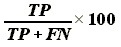
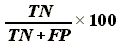
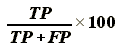
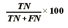
| POSITIVE TEST | NEGATIVE TEST | TOTALS | |
| PATIENTS WITH DISEASE | True Positive (TP) | False Negative (FN) | TP + FN
All patients with disease Sensitivity: |
| NORMALS | False Positive (FP) | True Negative (TN) | FP + TN
All patients without disease Specificity: |
| TOTALS | All patients testing
positive.
Positive Predictive Value:
|
All patients testing
negative.
Negative Predictive Value:
|
TP + TN + FP + FN
All patients tested. |
SENSITIVITY and SPECIFICITY: First do a test with high sensitivity, to screen the general population. Then, if that comes back positive, do a test with high specificity to confirm that the patient actually has disease.
Erythrocyte Sedimentation Rate (ESR): A high ESR occurs when the body is infected or under stress, and the liver is releasing acute-phase proteins into the blood.
ACUTE-PHASE PROTEINS: Proteins released by the liver when the body is under stress.
PROTEIN ELECTROPHORESIS: alpha1, alpha2, beta, and gamma zones all have distinct proteins.
GAMMOPATHIES:
PLASMA ENZYMES:
PROTEINURIA: Can be caused by three mechanisms:
TOTAL BODY WATER:
POTASSIUM: Reference range 3.5 - 5.0 mEq / L
SODIUM: Reference range 135 - 146 mEq / L
DEHYDRATION:
EDEMA:
OSMOLALITY: Normal value is about 289 mOsm.
ACID-BASE:
ANION GAP: Essentially, the difference between between the concentrations of cations (Na+ primarily) and anions (Cl-, HCO3-) in the blood.
NORMAL VALUES of ARTERIAL BLOOD-GASES:
| Item | Value |
| pH | 7.4 |
| [HCO3-] | 22 - 28 mEq / L |
| PaCO2 | 33 - 44 mEq / L |
| PaO2 | 90 - 100 mEq / L |
ACID-BASE CASE STUDIES:
| Case # | pCO2 | PO2 | HCO3- | pH | Explanation |
| Case 1 | 70 | low | 27 | 7.2 | Acute Barbiturate Overdose. PCO2 is high ------> respiratory acidosis from hypoventilation. It is uncompensated because the HCO3- is normal and the pH is low. |
| Case 2 | 70 | 100 | 12 | 7.0 | Code Arrest. High PCO2 ------> respiratory acidosis. Also, low HCO3- ------> metabolic acidosis. It's a mixed disorder. |
| Case 3 | 59 | 50 | 31 | 7.34 | COPD. Partially compensated respiratory acidosis. High PCO2, high HCO3- (metabolic alkalosis) in compensation, near normal but slightly low pH. |
| Case 4 | 29 | 100 | 22 | 7.50 | Hyperventilation. Uncompensated respiratory alkalosis. |
| Case 5 | 50 | 80 | 12 | 7.27 | Chronic Renal Failure. Patient shows partially compensated metabolic acidosis with high anion gap. Patient can't excrete all the acid he is creating. |
| Case 6 | 50 | 80 | 42 | 7.52 | Diuretics in a non-smoking female. Metabolic Alkalosis (high HCO3-) with partially compensated respiratory acidosis (low PCO2). |
| Case 7 | 62 | 50 | 36 | 7.37 | COPD, loop diuretic. Mixed disorder. Respiratory acidosis from COPD, and metabolic alkalosis from loop diuretic. The pH is near normal but it should not be called compensated, because full compensation never occurs, and the pH is the result of two unrelated processes. |
CLINICAL BIOCHEMISTRY CASE STUDIES:
| Case | Pertinent Lab Values | Explanation | |
| 1 | Potassium Lab Error, Addison's Disease | High K+
High Urea Low Na+ |
K+ was high becuase of partial hemolysis of blood, because blood was aged. Labs could indicate Addison's Disease, but they need to be retaken. |
| 2 | Potassium Lab Error | High K+ | K+ of 45 is incompatible with life. |
| 3 | IDDM | Glucose tolerance test: young kid most likely has a transitory hyperglycemia, because he just ate. Next day glucose is normal | |
| 4 | Starvation, Dehydration | ICF and ECF will shrink to the same extent.
Drink seawater: death due to hypernatremia, diarrhea from magnesium in the sea-water. | |
| 5 | Dehydration | High Na+, high Cl-
High urea (pre-renal failure) Low HCO3- (acidosis) |
Man lost pure water ------> dehydration with
hypernatremia. He had hypotension, high pulse.
Pre-renal failure: Due to inadequate perfusion of kidneys; uremia (high urea) is more prominent than high creatinine. |
| 6 | Paraneoplastic SIADH | Low serum osmolality,
low urine osmolality.
Low Na+, low Cl- High K+ (aldosterone is not being secreted at all) |
Differential should include Diabetic Ketoacidosis. |
| 7 | Dehydration | High urea
All electrolytes are low. Low HCO3-, acidosis. |
Uremia: pre-renal failure due to hypotension.
These labs would not be found in end-stage kidney failure. |
| 8 | Injury with Lactic Acidosis | High Na+
High K+ Low HCO3-, metabolic acidosis |
Hyperkalemia is often associated with metabolic
acidosis.
Give calcium chloride immediately to prevent arrhythmias associated with the hyperkalemia. |
| 9 | Volume depletion after surgery | Low Na+ | Her sodium was depleted from surgery. Her responses to the low sodium included all things except reduced GFR. |
| 10 | Creatinine Clearance | Erroneous collection of urine is most common mistake in measuring creatinine. | |
| 11 | Diabetes Insipidus | Normal electrolytes
(more or less).
High serum osmolality. Low urine osmolality. |
Lack of ADH. ADH effects osmolality and plasma volume, but not electrolyte balance. |
| 12 | Septicemia with acidosis, pre-renal failure | High K+
High urea High serum osmolality |
High K+ is associated with acidosis. Renal disturbance is due to pre-renal failure. Serum urea is also increased because patient is in a state of excessive catabolism. |
| 13 | Renal Osteodystrophy | High urea, high creatinine | Chronic renal failure ------> low, calcium and Vitamin-D ------> high PTH. |
| 14 | Compensated Metabolic Alkalosis | High HCO3- (alkalosis),
low PCO2 (compensatory acidosis).
pH is high, but variable. Low K+ |
Patient had severe vomiting, and later had shallow
respirations.
Low K+ is associated with alkalosis. |
| 15 | Respiratory Acidosis | High PCO2 (respiratory
acidosis).
High HCO3- (compensatory alkalosis) |
|
| 16 | Compensated Respiratory Acidosis | High PCO2 (respiratory
acidosis).
High HCO3- (compensatory alkalosis) |
pH is closer to normal, hence compensated. |
| 17 | Diuretic-induced hypokalemia with Metabolic Alkalosis | Low K+
High HCO3- |
|
| 18 | Membranous Nephropathy, Albuminuria | Albuminuria | |
| 19 | Multiple Myeloma | Hypercalcemia | |
| 20 | Myocardial Infarct | Increased CK, AST, LDH. | Creatinine Kinase MB (CK-MB) is most useful isoenzyme for diagnosis. |
| 21 | Metastatic Breast Cancer | High Alk.Phos, AST,
ALT.
Normal albumin |
Cancer metastases to bone. |
| 22 | Obstructive Jaundice, caused by Carcinoma of Head of Pancreas | Very high alk.phos, indicated of cholestasis.
High AST and ALT High Bilirubin |
|
| 23 | Acute Viral Hepatitis | Very high AST and
ALT
Moderate Alk.Phos. High gamma-GT |
Dark color of urine is due to conjugated bilirubin.
Patient should recover from the hepatitis without consequences. |
| 24 | NIDDM | glucose tolerance test | |
| 25 | Diabetic Ketacidosis | Odor on breath | |
| 26 | Nocturnal Hypoglycemia in a Diabetic | Low blood sugar at night after taking insulin. | Measuring blood sugar during a hypoglycemia
attack isn't practical. Can measure catecholamines in the blood to estabolish diagnosis.
Treatment: adjust insulin levels. |
| 27 | Osteomalacia | Low Ca+2
Low adjusted Ca+2 |
High alk.phos. would be found if ordered, to establish diagnosis. |
| 28 | Paraneoplastic Hypercalcemia | High Ca+2
Low phosphate Normal PTH |
Normal PTH was found on further investigation, so they took X-rays looking for metastases. |
| 29 | Hypomagnesemia with scondary Hypoparathyroidism. | Low Mg+2
Low PTH secondarily |
Mg+2 is required for PTH secretion! |
| 30 | Paget's Disease of Bone | High alk.phos. | |
| 31 | Lactotrope Adenoma with Pan-hypopituitarism | High Prolactin
The rest of the pituitary hormones are low |
Compression atrophy of the rest of the pituitary. |
| 32 | Possible Growth Deficiency | Repeat test. GH levels can fluctuate, and erroneous results can happen after a single random measurement. | |
| 33 | Cystic Cold Thyroid Nodule in woman on ERT. | High T4
Low TSH |
Taking estrogen ------> TBG is higher ------>
T4 baseline must be higher to compensate for the
increased TBG.
Perform fine-needle aspiration biopsy to evaluate the nodule. |
| 34 | Hypothyroidism | ||
| 35 | Thyrotoxicosis | Order free T3 and T4 tests to evaluate status. | |
| 36 | Acute Adrenal Cortical Failure | Low Na+, High K+
Hypotension Acidosis |
Low Na+ and high K+ result from no aldosterone.
Acidosis is secondary to the hyperkalemia. Give ACTH (Synachten) test to confirm diagnosis. |
| 37 | Auto-immune Adrenalitis (Addison's Disease) | Low Na+, High K+
Hypotension Acidosis |
|
| 38 | ACTH-Secreting Carcinoma of Lung, Cushing's Disease | Carcinoid tumor. | |
| 39 | Polycystic Ovary Syndrome | High testosterone
High LH, low FSH |
|
| 40 | Chronic Malnutrition | Vitamin-K malabsorption | |
| 41 | Pernicious Anemia with Hypothyroidism | ||
| 42 | Total Parenteral Nutrition, secondary hyperglycemia | High blood sugar | Can see hyperglycemia in patients who are on TPN, due to poor or no stimulation of insulin release. |
| 43 | MVA with tissue injury | High K+ | High K+ is released from tissues, from tissue injury.
Measure creatinine kinase to document muscle cell necrosis (rhabdomyolsysis). |
| 44 | Osteomalacia | High alk.phos.
Low Ca+2, low Vit-D |
Most likely caused y malnutrition, or malabsorption of Vitamin-D. |
| 45 | Iron-Deficiency Anemia | Low Fe+2
Low transferrin saturation (high binding capacity) Low ferritin. |
|
| 46 | Wilson's Disease | Liver failure. | |
| 47 | Digoxin Toxicity, Renal Failure | Patient had elevated serum urea due to pre-renal failure, secondary to heart failure. | |
| 48 | Salicylate Poisoning | Low HCO3-
High anion gap |
Metabolic Acidosis with Respiratory Alkalosis.
Anion gap is increased because it is metabolic acidosis. |
| 49 | Lead Poisoning | Measure protoporphyrin in blood cells to confirm diagnosis. | |
| 50 | Alcoholism | There is no lab test that is specific for alcoholism. gamma-GT comes close but is not diagnostic. | |
| 51 | Diabetic Hypoglycemia after drinking alcohol | Patient was hypoglycemia, due to mixing alcohol with insulin. Treat with IV glucose. | |
| 52 | Hyperlipidemia | Low electrolytes
High amylase High triglycerides |
Pseudohyponatremia: Low Na+ due to abnormally low water-content of plasma (i.e. plasma
had way too much lipid in it).
Genetic disorder involves Apolipoprotein-B Patient is at risking of forming a volvulus. |
| 53 | Obesity, hyperlipidemia, NIDDM, Alcohol | High cholesterol, lipids
High glucose High gamma-GT |
Treat with dietary measures. Man is at increased risk for coronary artery disease. |
| 54 | Heterozygous Familial Hypercholesterolemia | High fasting cholesterol.
Normal lipids Low HDL |
Hypercholesterolemia is also found in patients with Hypothyroidism. |
| 55 | Pheochromocytoma | VMA in urine.
HTN |
|
| 56 | ACTH-secreting tumor, Cushing's Disease | High HCO3-
Low K+, High Na+ High creatinine |
Metabolic Alkalosis secondary to hypokalemia,
from increased aldosterone activity.
Probably comes from oat-cell carcinoma of lung. |
| 57 | Alcoholic Liver Disease, Hepatoma | High liver enzymes
High gamma-GT |
alpha-Fetoprotein was normal in this case (it's
usually elevated)
Can also measure Carcinoembryonic Antigen (CEA) |
| 58 | Thyroid Carcinoma | Severe headache
High Ca+2 |
|
| 59 | Septic Arthritis posing as Gout | Uric acid came back normal.
Give antibiotics to treat septic arthritis. | |
| 60 | Hemolysis, Tissue Damage | High LDH, high CK
Low haptoglobin |
LDH, CK = damage to: muscle, liver, or erythrocytes. |
| 61 | Cystic Fibrosis | High Cl- in sweat | |
| 62 | Rh-Incompatibility Disease | Measure bilirubin in amniotic fluid to diagnose erythroblastosis fetalis. High bilirubin would indicate hemolysis in the fetal blood. | |
| 63 | Pre-Eclampsia | Progressive albuminuria, HTN | |
| 64 | Cretinism | Baby came back normal. TSH must be above 100 before follow-up test is required. | |
| 65 | IRDS in premature infant |