Liposomes
Use, formation, and important surface considerations.
www.avantilipids.com/
Introduction
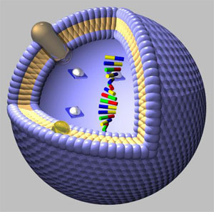
Liposomes are sphere-like vesicles composed of lipid molecules similar in composition to the cell membranes found in biological systems. They are of great interest to the pharmaceutical industry as drug-delivery vectors due to the fact that they can encapsulate hydrophobic compounds in the lipid bilayer, and at the same time hydrophilic compounds in their inner aqueous volume. Liposomes are being developed to target specific regions of tissue by the use of membrane bound antibodies that have antigenic specificity for certain cell types, and then fuse with them - delivering their bound cargo.
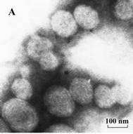
Electron Micrograph of Liposomes***
Formation of Liposomes
Liposomes can be formed by a process called mechanical dispersion or thin-film hydration. In this method, lipids are dried onto a solid support such as the side of a glass container and then dispersed by the addition of an aqueous medium, followed by shaking. When hydration occurs in the system, the lipids swell and peel off of the support in sheets to form multilamellar vesicles (liposomes). **
Applications***
Liposomes can be used as anticancer delivery agents primarily in the reduction of toxic effects from the injected drugs. These phospholipid vesicles can encapsulate both hydrophobic and hydrophilic drugs for delivery to specific sites. A key feature of liposome effectiveness is its ability to be biodegradable in vivo.
The following list summarizes the key advantages of liposomal drug delivery:
- solubilization – liposomes solubilize lipophilic drugs
- protection – encapsulated drugs are inaccessible to metabolized enzymes, also body components are not directly exposed to the full dose of the drug
- duration – liposomes prolong drug action by slowly releasing the drug in the body
- directing potential – liposomes can be used with targeting options to change the distribution throughout the body
- internalization – liposomes can be endocytosed or phagocytosed by cells
- amplification – liposomes can be used as adjuvants in vaccine formations
Zeta Potential Measurements of Liposomal Dispersions
The determination of the zeta potential of liposomes can be achieved by using a liposomal suspension diluted with distilled water, then subjected to photon correlation spectroscopy and laser Doppler anemometry.
Photon correlation spectroscopy is applicable to particles suspended in a liquid, which are in a state of random movement due to Brownian Motion. The pace of the movement is inversely proportional to particle size (the smaller the particles are, the faster they move, or diffuse), and the pace can be detected by analyzing the time dependence of light intensity fluctuations scattered from the particles when they are illuminated with a laser beam.
Laser Doppler
Anemometry measures velocities of small particles in flows. The technique is
based on the measurement of laser light scattered by particles that pass through
a series of interference fringes (a pattern of light and dark surfaces). The
scattered laser light oscillates with a specific frequency that is related to
the velocity of the particles. Particle velocities are usually expressed as
electrophoretic mobilities. The electrophoretic mobility (EM) of a particle is
defined as follows:
EM=u/field strength
The zeta potential (ZP) can be calculated by application of the Smoluchowski
equation
ZP=4 ηu/e
where η is the viscosity of the medium, e is the dielectric constant and u is
the particle velocity, respectively.
Surface Chemistry Considerations: Stability of Liposome Dispersions (Colloidal Stabilization by Hydration Force)*
The following is a description of the work performed by Sabin, Prieto, Ruso, Hidalgo-Alvarez, and Sarmiento to explain the role of hydration in the stabilization of liposomal dispersions. Their whole work and references may be found here
Liposomes have been described as associating colloids, formed of amphoteric lipids that self-assemble into spherical, closed structures. Liposome dispersions however, are not thermodynamically stable. There is a tendency for the system to lower its energy by reducing its interfacial area. The tendency of liposome dispersions to aggregate is due to the attractive forces of van der waals interactions. To understand these interactions, and effectively manage them is critical to the use of liposomes as vehicles for drugs, enzymes, or genetic material.
The DLVO (Derjaguin, Landau, Verwey, Overbeek) theory is typically used to predict colloid stability. The application of DLVO theory to liposomal dispersions is somewhat limited. This is because a critical phenomenon has been overlooked in applying DLVO to liposomal dispersions: the hydration sphere of ions adsorbed onto the lipid bilayer of the liposomes. Also, classical DLVO theory was developed for rigid colloids, which liposomes are not. The lack in the DLVO theory was realized once atomic force microscopy allowed for the measurement of surface interactions at very short separations.
Marcelja and Radic consider a force associated with the nature of the solvent (in this case water). Marcelja and Radic consider that the bilayer surface causes the water molecules to orient in contrast with the surface. The orientation is propagated in a damped way out of the surface through successive layers of interaction.
At low salt concentrations, repulsion due to the double layer is the dominant surface force. As the concentration of salt increases, the hydration force due to adsorbed ions becomes the dominant repulsive force. Below is an image displaying an ion adsorbed onto the outer layer of the lipid bilayer of a liposome.
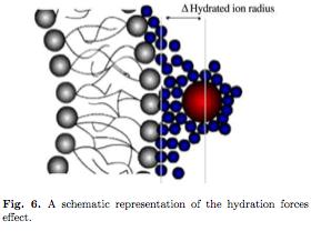
Hydration forces clarify the stability of systems such as liposomal dispersions where DLVO theory is lacking. Sabin, Prieto, Ruso et al. have attempted to modify DLVO theory to take into account the effect of hydrated ions adsorbed to liposomal surfaces.
Preparation and Zeta-Potential Measurements
Liposomes were prepared by the thin-film hydration method, and the electrophoretic mobilities of the liposomes were measured to determine zeta-potential. The zeta-potential was calculated using the Henry correction of Smoluchowski's equation:
Eq. 1

Where ε0 is the permittivity of free space. εr is the relative permittivity, a is the radius of the liposome, 1/ κ is the Debye length and η is the viscosity of water, μ is the velocity of the particle under an electric field. f(κa) is dependent upon particle morphology and the ionic strength and is found by equation 2.
Eq. 2
![]()
Equation 2 is valid for values of κa greater than 10.
DLVO and Beyond
In DLVO, the potential of interaction between two particles is the sum of an attractive force and a repulsive force. The attractive force being van der Waals interactions, and the repulsive force is due to electrostatics. In general this gives us equation 3.
Eq. 3
![]()
Where Vt is the total interactive force, Va is attractive forces, and Vr is repulsive forces.
To find Va we use Hamaker's equation for the case of two spheres of equal radius a and thickness d. (Equation 4)
Eq. 4
![]()
Where A is the Hamaker constant, and x is the distance between the two particles.
We find the repulsive force Vr using equation 5.
Eq. 5
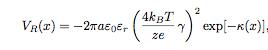
Where kB is the Boltzmann constant, T is temperature in Kelvins and κ is found by Equation 6.
Eq. 6
![]()
Where cs is the ionic concentration, e is elementary charge, zi is the valence of the adsorbed ions, and Na is Avogadro's number and γ is given by Equation 7.
Eq. 7
![]()
Where ξ is the zeta-potential.
The zeta-potential is determined at a location a small distance from the bilayer surface called the surface of shear. The zeta potential magnitude is reduced at this distance depending strongly on the Debye length. The distance to the shear plane is highly dependent upon solution ionic strength and described by the Gouy-Chapman-Stern theory. With the high molarities used in this study, the authors assume that the membrane potential is screened only by monovalent ions, and that the membrane potential and zeta potential are one and the same. The zeta-potential can be measured as a function of monovalent ion concentration and the results are in the figure below.
Fig. 3
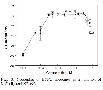
In the above figure EYPC stands for (Egg Yolk Phosphatidylcholine) which is what the liposomes are formed of. As can be seen in figure 3, zeta potential is greatly quenched by the concentration of monovalent cations.
Sabin et al use a Hamaker constant in the range of 3.75-5.48 x 10^-20 J in equations 4 and 5 to obtain the classical DLVO curve for different salt concentrations as a function of distance between liposomes.
Classical DLVO for Liposomal Dispersions
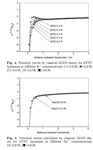
If classical DLVO theory is correct for this dispersion of liposomes, then we would expect a rapid aggregation due to van der Walls attraction forces. This is not observed, therefore the DLVO theory is lacking in some way that will shortly be improved upon. Sabin et al propose that a 'hydration force' is responsible for the stability of the dispersion. The authors add a correction factor to the DLVO equation to take into account the hydrated ions absorbed onto the surface of the liposomes: This gives us equation 8.
Eq. 8
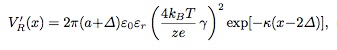
Where Δ is the hydrated ion radius of the absorbed ion. (0.36 nm for Na + and 0.33 nm for K +). When the hydration forces are taken into account, a repulsion barrier appears in the potential curves of the modified DLVO curve. Sabin et al propose that this is the reason for the stability observed in their EYPC liposome dispersions. This provides us with the following figures.
Modified DLVO for Liposomal Dispersions
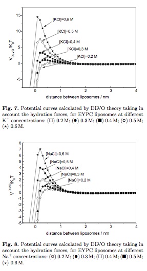
Sabin et al. conclude that EYPC liposomes are stabilized due to the hydration force of repulsion, caused by the absorption of monovalent cations to the surface of the liposomes, and have expanded DLVO theory to include hydration forces as a stabilizing factor for liposomes in colloid science.
*All images and equations in this section are taken from Sabin et al. Their full work may be found by clicking the link at the beginning of the section.
** Liposomes, a Practical Approach, IRL Press 1990, Pg 36
*** Zheng-hong Wu, Qi-neng Ping, Yi Wei, Jia-ming Lai. Hypoglycemic efficacy of chitosan-coated insulin liposomes after oral administration in mice. Acta Pharmacol Sin, July 2004.