
HIV picture / photo
 The
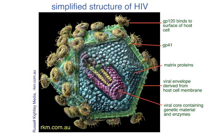
structure of the Human Immunodeficiency Virus (HIV) is relatively simple. The viral
contains the genome, or genetic
material, which is accompanied by several
essential to successful reproduction, including protease
(p9) [for "protein 9"], reverse
transcriptase & RNAse (p66), and integrase
(p32). This material is held within the
, a
single layer of structural proteins (p24).
Surrounding this in turn is the matrix protein membrane, composed of additional
structural proteins (p17). Finally, encasing all these elements, is the viral
which is composed of lipids taken
from the host cell (usually an human immune cell
such as a T-Lymphocyte or macrophage). Protruding through the lipid membrane are
numerous spikes of envelope
which are used by the virus to facilitate attachment
to the host cell via various cell surface receptors. The envelope
glycoproteins have two separate parts: the surface glycoprotein (SU), designated
gp120, and the transmembrane glycoprotein (TM), designated gp41.
Please note that these numbers represent the approximate atomic weight of these
proteins, expressed in daltons. The sum of these two weights (120
+ 41) equals roughly 160 daltons, hence the desgnation of the combined
envelope glycoprotein as gp160.
The
structure of the Human Immunodeficiency Virus (HIV) is relatively simple. The viral
contains the genome, or genetic
material, which is accompanied by several
essential to successful reproduction, including protease
(p9) [for "protein 9"], reverse
transcriptase & RNAse (p66), and integrase
(p32). This material is held within the
, a
single layer of structural proteins (p24).
Surrounding this in turn is the matrix protein membrane, composed of additional
structural proteins (p17). Finally, encasing all these elements, is the viral
which is composed of lipids taken
from the host cell (usually an human immune cell
such as a T-Lymphocyte or macrophage). Protruding through the lipid membrane are
numerous spikes of envelope
which are used by the virus to facilitate attachment
to the host cell via various cell surface receptors. The envelope
glycoproteins have two separate parts: the surface glycoprotein (SU), designated
gp120, and the transmembrane glycoprotein (TM), designated gp41.
Please note that these numbers represent the approximate atomic weight of these
proteins, expressed in daltons. The sum of these two weights (120
+ 41) equals roughly 160 daltons, hence the desgnation of the combined
envelope glycoprotein as gp160.
Image of the structure of a mature HIV virion
3D HIV structure
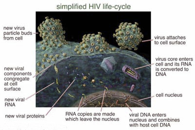
Simplified HIV life-cycle
The initial phase of HIV infection is binding. The virus attaches to the host cell, in this case a CD4 Lymphocyte (or T-4 cell), by binding to the various cell surface receptors available to it. These include the well-known surface marker and a number of chemokine receptors that have recently been identified as essential co-receptors in HIV infection. In T-cells, the CC-chemokine receptors are used; in macrophages, the b-chemokine receptors are used. With this discovery comes the possibility of new drug therapies to block attachment through these essential co-receptors. [Source: science-mag.aaas.org/science]
The three major HIV enzymes present the best targets yet for new drug therapies. The reverse transcriptase inhibitors, such as the nucleoside analogue and the non-nucleoside RT inhibitors were the first drugs developed against HIV, and have been moderately successful in achieving limited suppression of viral replication. The protease inhibitors such as the drugs ritonavir, indinavir and nelfinavir, has been far more effective at suppression of viral replication, achieving almost total suppression in many patients taking these drugs
However, the development of drugs
to suppress viral
enzymes and
genes has not been without its failures. Past efforts to create drugs that
inhibited TAT
(a protein
involved in upregulating viral reproduction) were a dismal
failure. It
is hoped that next-generation
therapies such as
the integrase
inhibitors, already
in early development, may be more effective than even the protease inhibitors.
[Source: [email protected]]
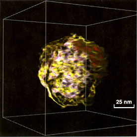
Immature HIV, human immunodeficiency virus ( ), low-pass filtered to 6 nm.(electron tomography). Electron tomography consists of scanning of micrographs and the resulting pictures must be aligned with respect to one another. Scaling, rotations, and translation of origin have all to be checked because of the variations in them from one picture to another introduced by the manual microscopy and scanning.
HIV,
human immunodeficiency virus (Retroviridae): Spherical, enveloped i.e. membrane
bound particles (100-130 nm i diameter). The genome consists of two (diploid)
identical ss-RNA strands (plus t-RNA from the host). As seen by
thin-sectioning-techniques, the envelope of immature
virions is lined with a 20-25 nm thick
submembraneous layer of precursor (gag) proteins and
the central area
looks empty. Mature
virions have in the
center a characteristic conical
core consisting of the
viral RNA complexed with viral protein and
the submembraneous layer is
very thin. Platina-carbon shadowing
techniques have revealed globular knobs (12-14 nm in diameter and 10 nm high)
formed by clusters of glycoproteins. The knobs
have the
; believed to
make the viruses more resistant to immunological
attacks. However, the knobs are also sensitive to preparative conditions. They
are not easily distinguished e.g. in thin sections. In negative-staining with
uranyl acetate, merely triangular shaped protrusions are detectable, interpreted
that the lower part of the knobs is triangular
while the top is globular and
covered by stain i.e. making them less visible
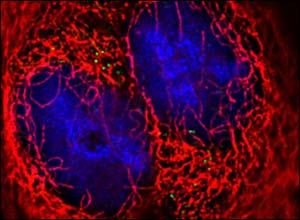
The pictures (fluorescent microscopic) offer proof that the virus uses the cell's own machinery to be dragged inside the cell..The study, by the University of Illinois at Chicago, reveals how HIV "hitches a ride" aboard a cell protein called as it makes it way up tiny microtubules into the cell itself. The tubules lead all the way to the cell's nucleus, where the virus can begin to start replicating by harnessing the cell's own ability to copy its genetic code. Virus particles are only approximately 12 millionths of a centimetre in diameter, yet the University of Illinois team managed to produce pictures of them as they headed for their destination. They did this by attaching green fluorescent proteins from jellyfish to them, then shining a blue light on them to make them glow. Each HIV particle appears as a green dot on the background of the red tubules, which have also been marked with another fluorescent protein. Time lapse photographs were taken under the microscope at 15 second intervals to reveal the virus' steady progress towards the centre of the cell. Professor David McDonald, one of the scientists leading the project, said: "They don't make a beeline for the nucleus." Their progress is somewhat halting. They appear to from one microtubule to another, moving in a jagged path, even sometimes moving backward, but they eventually reach their destination." Professor Thomas Hope, another scientist involved in the project, said: "We hope this basic research will one day lead to new targets for drug therapy in the longstanding battle against Aids." The pictures were published in the Journal of Cell Biology.
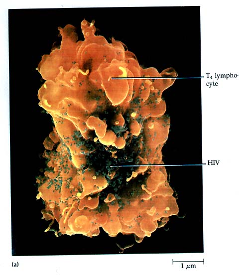
This is a scanning electron micrograph show HIV (dark green area) surrounding the T4 lymphocyte. The source of this picture was Microbiology, fourth edition, by Tortora, Funke, and Case, The Benjamin/Cummings Publishing Company, Inc, page 3


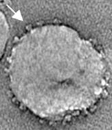
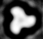
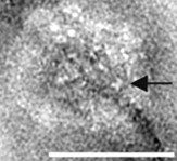
The pictures (electron microscope) on the 1st and 2nd picture show engineered SIV [simian (monkey) immunodeficiency virus] expressing high levels of the surface protein gp120 (indicated by arrows) The -like shape (3rd picture) of the gp 120 can be clearly seen . The final picture shows a wild type HIV particle, showing the much lower density of gp120 (which came as a surprise; most researchers thought it would look like the SIV pictures). The white bar in the HIV picture is 100 nm, about 1/500th the thickness of a human hair; the same scale applies to the SIV pictures.
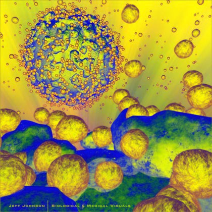
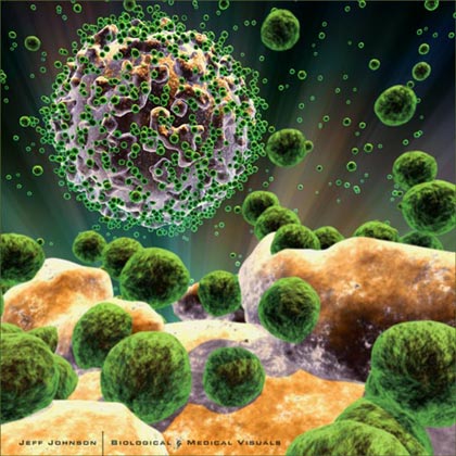
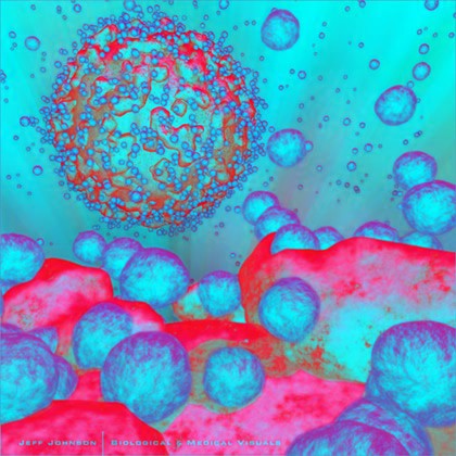
Graphical 3D HIV attacking T4 lymphocyte

![]()

