| Photomicrographs of Coals |
| An ornate megaspore in white light (left) and in fluorescence light (right). |
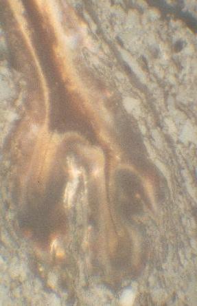
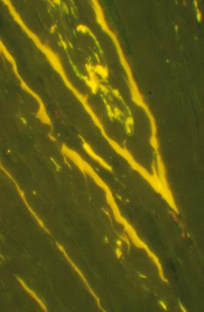
| A cross section through a pinus needle, showing the outerlayer cuticle with some internal cell structure higlighted by resinite, in white light (left) and in fluorescence light (right). |
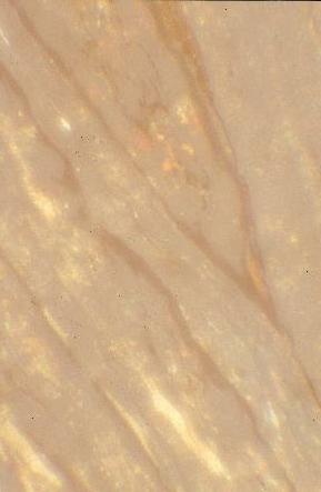
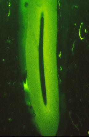
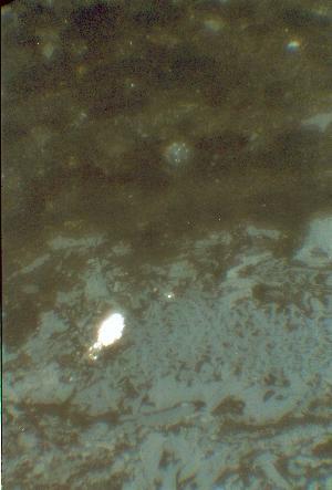
| A seed showing a low level flourescence, this is one of the first seeds to be seen in the fossil record; in white light (left) and in fluorescence light (right). |
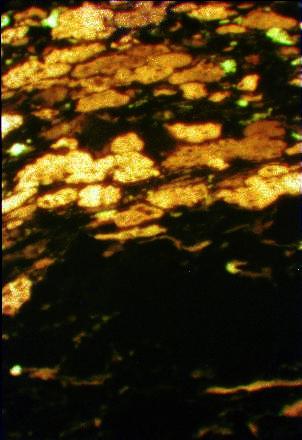
| Highly fluorescing algal colonies (alginite) in the top half of the photo and a low-reflecting semi-fusinite band below; in white light (left) and in fluorescence light (right). |
| ...the following photomicrographs come from the Mid-Cretaceous, Lloydminster and Cummings coal seams, Alberta. |
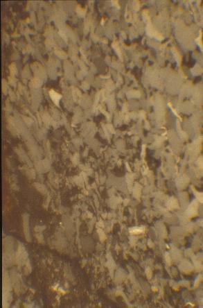
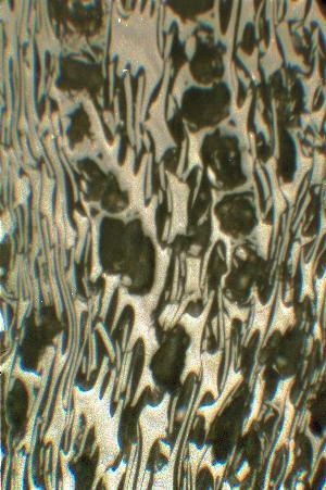
| Inertodetrinite that shows micrograded bedding. The grain size of the detrital coal increases from bottom left to top right. |
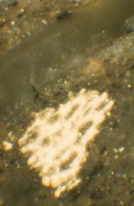
| Highly reflecting fusinite showing original cell lumens (windows). This inertinite was probably formed by a palaeofire. |
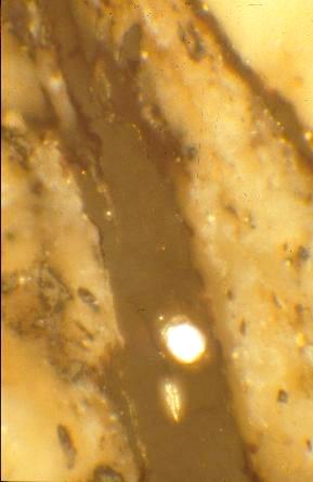
| Pyrite (FeS2) that has completely replaced fusinite, in a shale. |
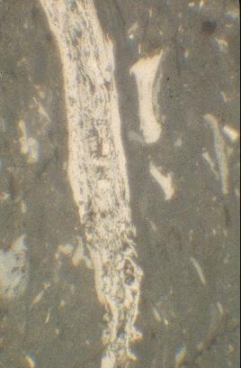
| Telovitrinite rootlet in a shale. Note framboidal pyrite contained with rootlet. Sulphate-reducing bacteria use organic matter with pyrite beign the by-product. |
| Cross-section through leaf that has been oxidised to semi-fusinite. |
 |
 |
 |
 |
 |
 |
 |
 |
 |
 |
 |
 |