 |
|
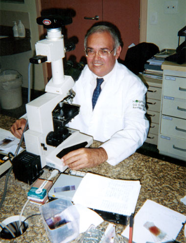
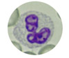
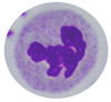
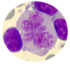
Over the course of his 25-year career, Brazilian Pathologist, Nivaldo Medeiros, M.D. has discovered a different set of patterns in the slides that pass under his microscope every day. During his work at The University of São Paulo School of Medicine's Clinical Hospital and The Cancer Hospital, both in São Paulo, Dr. Medeiros has found, photographed and collected cellular patterns of leukocytes, erythrocytes and platelets that bear a striking resemblance to different animals, kitchen utensils, and people engaged in various activities. His collection, which today numbers some 300 images, is looked upon with a good deal of curiosity by his colleagues. "Anyone could see these patterns, if they took the time to look for them. They're not that hard to find, nor do they represent especially rare pathologies" says Dr. Medeiros. "More often than not, they're simply random groups of cells, without any particular reason for their aggregation," he continues. To find these curious patterns, Dr. Medeiros typically uses a magnification of 1000X with a Leishman panchromic (panoptical) stain. As you can see from these photos from Dr. Medeiros' collection, there a great many of these patterns that form everyday things that most pathologists never bother to notice. |
|
|


|
The
reproduction or use of these photos is strictly prohibited without the
author's permission. All rights reserved.
|
|
In addition to his "hobby" collection, Dr. Medeiros has also amassed approximately 4,500 photos that cover much of the gamut of hematological pathologies. His goal is to grow this collection to 7,000 photos, when he will then decide the final destiny of his long and exhaustive work. About
the author:
Nivaldo Medeiros, MD Former Director of the Hematology and Cytology
Service of the Central Laboratory of the University of São Paulo School
of Medicine.
|