Projects
|
Human endogenous retrovirus (HERV) distribution in non-human primates
|
|
|
I am interested in the evolution of HERVs in general and more specifically, when they entered and expanded copy number in the genome of various primates. Did they all enter at once? Are there specific points in primate evolutionary history during which HERVs were particularly transpositionally active? To address this, we are analyzing a large sampling of non-human primates to examine the distribution of all classes of HERV's using quantitative PCR and HERV specific DNA microarrays (Seifarth et al. 2003). The results are intriguing and suggest a major event in the evolution of HERVs occurred after New World Monkeys separated from Old World monkeys. Considering that medically relevant HERV sequences have been discovered including some with potential roles in disease, the evolutionary history of each HERV class could help guide researchers to potentially medically relevant HERVs.
|
References:
Seifarth, W., Spiess, B., Zeilfelder, U., Speth, C., Hehlmann, R., and Leib-Mösch, C. (2003) Assessment of retroviral activity using a universal retrovirus chip. J. Virol. Methods. 112: 79-91.
|
Endogenous retroviruses as co-factors in prion disease pathogenesis
|

|
The project to investigate the potential role of endogenous retroviruses in prion disease pathogenesis is part of the FORPRION funded group of projects dedicated to understanding prions and the diseases they cause. For information about this project see here.
|
Identification of changes in gene expression in response to prion infection
|

|
In collaboration with the Expression Profiling Group of the Institute for Experimental Genetics at the GSF and with the prion research group of Professor Hermann Schätzl at the Institute for Clinical Virology of the Technical University of Munich, we are searching for co-factors in scrapie pathogenesis in mouse neuronal cells. Using well established cell lines that can be persistently infected we have screened for differences in gene expression in over 22,000 expressed sequences using DNA microarrays to identify up and downregulated genes. We have also employed quantitative PCR for selected genes of interest. The results have yielded some intriguing candidates including several genes known to be altered in regulation in other neurodegenerative diseases. Continued work on these and additional cell lines should provide a clearer understanding of the influence of scrapie on the biology of the cells it affects.
|
Ancient DNA
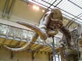
Woolly mammoth phylogenetics
|
|
|
Placing the extinct woolly mammoth (Mammuthus primigenius) in the elephant family tree has been plagued with difficulty. Morphological analysis suggests that mammoths were more closely related to Asian elephants (Elephas maximus) than to either of the extant African elephant species (Loxodonta africana and Loxodonta cyclotis). However, many of the characters used are subject to convergent evolution and may give a confusing picture. Molecular studies using mitochondrial gene sequences have also been problematic. For one thing, the elephant genome is littered with nuclear copies of mitochondrial genes (Numts) that can cause a great deal of confusion when these sequences are used for phylogenetics (Greenwood and Pääbo, 1999). Another problem is that different researchers using mitochondrial sequences have come to opposite conclusions regarding mammoth phylogeny (Greenwood 2001). This may be a result of unintentional inclusion of Numts, sequence artifacts that frequently occur during PCR from ancient DNA (Pääbo et al. 1990), or the limitations of using a maternally inherited locus such as the mitochondrial genome.
In collaboration with Dr. Cristian Capelli of the Istituto di Medicina Legale of the Universit96a Cattolica del S. Cuore in Rome, Dr. Alfred Roca of the NIH, in Bethesda, Dr. Ross MacPhee of the American Museum of Natural History in New York, and the company Medigenomix (www.medigenomix.de), we are trying to resolve the issue of the woolly mammoth phylogenetic relationship with extant elephants using nuclear DNA sequences. We have demonstrated previously that characterizing multicopy and single copy DNA is possible (Greenwood et al. 1999, Greenwood et al. 2001). We have now extended this work and have sequenced several nuclear gene sequences from different mammoths and hope to have the answer soon.
|
References:
Greenwood, A.D. and Pääbo, S. (1999) Nuclear insertion sequences of mitochondrial DNA predominate in hair but not in blood of elephants. Molecular Ecology 8: 133-137.
Greenwood, A.D., Capelli, C., Possnert, G., and Pääbo, S. (1999) Nuclear DNA sequences from late Pleistocene megafauna. Molecular Biology and Evolution 16(11): 1466-1473.
Greenwood, A.D., Lee, F., Capelli, C., DeSalle, R., Tikhonov, A., Marx, P.A., and MacPhee, R.D.E. (2001) Evolution of endogenous retrovirus-like elements of the woolly mammoth (Mammuthus primigenius) and its relatives. Molecular Biology and Evolution 18(5): 840-847.
Greenwood, A.D. (2001) Mammoth biology: Biomolecules, phylogeny, Numts, nuclear DNA, and the biology of an extinct species. Ancient Biomolecules 3: 255-266.
Pääbo, S., Irwin, DM., and Wilson, A.C. (1990) DNA damage promotes jumping between templates during enzymatic amplification. J. Biol. Chem. 265: 4718-4721.
|
Ancient DNA
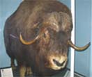
The genetic consequences of end-Pleistocene extinctions: Muskoxen as a test case
|
|
|
At the end of the Pleistocene (about 10,000 years ago), a mass extinction event occurred which primarily affected megafaunal mammals. This included such species as the woolly mammoth (except for a small population that survived a few thousand years longer), the woolly rhino, multiple groups of giant ground sloths, sabre tooth cats and many others. Some megafaunal species survived this event such as muskoxen (Ovibos moschatus). Or did they? Carbon dating of muskoxen bones from the Taimyr peninsula in Russia suggests that they disappeared at the end of the Pleistocene (MacPhee et al. 2002). They then reappeared several thousand years later only to disappear from the area completely about 2000 years ago. During this entire interval, they survived in parts of Canada and Alaska.
In collaboration with Dr. Ross MacPhee of the American Museum of Natural History, Medigenomix (www.medigenomix.de), and Scientific Research & Development GmbH, we have analyzed DNA from muskoxen bones from the Taimyr peninsula, Canada and other parts of Russia that both pre-date the end-Pleistocene extinctions and some which are much younger. The results show that before the end of the Pleistocene, a genetically distinct muskoxen group lived in Taimyr. All other muskoxen, including the younger Taimyr samples are virtually identical to modern muskoxen. Thus, it appears the end-Pleistocene extinctions were more severe than previously thought.
|
References:
Macphee, R.D.E., Tikhonov, A.N., Mol, D., de Marliave, C., van der Plicht, H., Greenwood, A.D., Flemming, C., and Agenbroad, L. (2002) Radiocarbon chronologies and extinction dynamics of the late Quaternary mammalian megafauna of the Taimyr peninsula, Russian Federation. J. of Arch. Sci. 29(9): 1017-1042.
|
|
|