Evaluate the state of Retinal Non Perfusion "If
more than 10 DD"
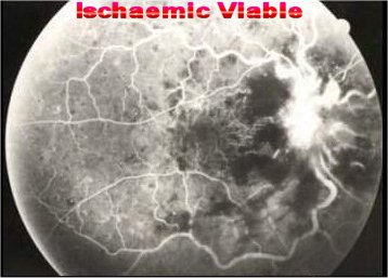
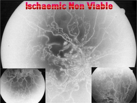
Evaluate the state of Reinal Tissue Viability "ERG"
|
Non Viable
|
Viable
|
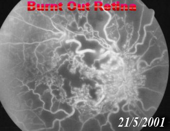
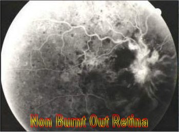
| Burnt Out Retina |
Non Burnt Out Retina |
| Less possible to complicate |
More Likely to complicate |
NVD / NVE
Neovascular Glaucoma "Gonio examination without dilatation" |
Evaluate Retinal Perfusion
after 6 months
FFA
|
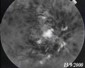
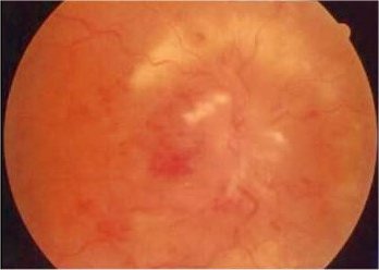
Persistent Macular Oedema with Cystoid
Formation
|
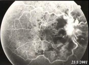
Capillary Non Perfusion involving FAZ
|
|
Grid Photo Coagulation
|
Nothing help
|
Follow "3-4 m" FFA to evaluate state of macula
|
oedema Improves
|
oedema persists
|
|
Keep Follow
|
Retreat Grid
|
|
|
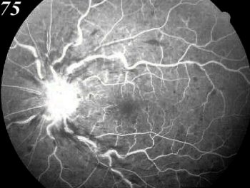
Area Of capillary Non
Perfusion > 5 DD
Start PRP to avoid Neovascular Glaucoma
|
Keep Follow for:
NVD / NVE
Neovascular Glaucoma "Gonio examination without dilatation"
Prophylactic Mild Scatter PRP may and may
not be
Better Follow/4m or apply if NVD or NVE appears |
|