Learning Visualized, on the Double
by Marcia Barinaga
The following has been edited by John Schmidt for the students in
the
Neurobiology course
he teaches. See the original article in Science
for the full article. Gerald
Edelman produced what is still the most complete theoretical outline
of how a brain creates a mind. As someone who was involved in the study
of cell adhesion molecules, he gave surprisingly little attention to the
idea of regulation of synaptic structure rather than simply regulation
of the function of existing synapses. This bias reflected the "conventional
wisdom" based on electrophysiology and gross brain anatomy that during
embryonic development the making and breaking of synaptic connections is
important, but in adult brains, all you need to do is regulate the function
of existing synapses. Fortunately, the tools are now becoming available
that allow us to visualize the rather subtle structural changes in synapses
that are involved in learning and memory.
As Marcia Barinaga introduces the situation, "Researchers have
long believed that when the brain learns, the synapses, the connections
between neurons, get stronger. For years, neuroscientists focused on chemical
changes that boost synapse strength, but more recent work suggests that
synapses change structurally, too." She points to a recent article in the
journal Nature by Dominique Muller that "reveals one dramatic change:
Some strengthened synapses actually double, with a second synapse quickly
forming right next to the one that was active". Muller has long been involved
with efforts
to link regulation of synaptic cell adhesion molecules (even Edelman's
favorite, NCAM) to learning and memory storage.
 The
method used was to stimulate the neurons of rat brain tissue slices to
produce a form of synapse strengthening called long-term potentiation (LTP),
which is the best studied physiological analog of learning in mammals.
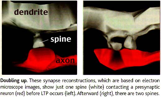
Electron microscopy was used to view synapses where LTP had occurred. The
trick was to take advantage of the fact that the activated synaptic spines
where LTP accurs contain higher levels of calcium ions. As Barinaga explains,
"Muller's team treated the brain slices with a chemical that precipitates
calcium, forming deposits that can be seen in the EM and serve as tags
for spines that had undergone LTP. One hour after inducing LTP, 20% of
the tagged synapses had double spines, both contacting the same presynaptic
neuron, a configuration that he very rarely saw in synapses that hadn't
undergone LTP. The authors conclude that LTP triggers 'a duplication of
the active synapse,' presumably strengthening it".
The
method used was to stimulate the neurons of rat brain tissue slices to
produce a form of synapse strengthening called long-term potentiation (LTP),
which is the best studied physiological analog of learning in mammals.
Electron microscopy was used to view synapses where LTP had occurred. The
trick was to take advantage of the fact that the activated synaptic spines
where LTP accurs contain higher levels of calcium ions. As Barinaga explains,
"Muller's team treated the brain slices with a chemical that precipitates
calcium, forming deposits that can be seen in the EM and serve as tags
for spines that had undergone LTP. One hour after inducing LTP, 20% of
the tagged synapses had double spines, both contacting the same presynaptic
neuron, a configuration that he very rarely saw in synapses that hadn't
undergone LTP. The authors conclude that LTP triggers 'a duplication of
the active synapse,' presumably strengthening it".
 Tobias
Bonhoeffer has been involved in studies of LTP. Barinaga interviewed
Bonhoeffer and reports that he views the work of Muller's lab as a "nice
addition" to the growing story of how synapses reshape when they strengthen.
Another report in Science from earlier this year by Bonhoeffer's
team and one led by Roberto
Malinow and Karel Svoboda at Cold Spring Harbor Laboratory on Long
Island used confocal microscopy of the synapses in living tissue to visualize
what looked like new spines popping out of neurons near strengthened synapses.
Tobias
Bonhoeffer has been involved in studies of LTP. Barinaga interviewed
Bonhoeffer and reports that he views the work of Muller's lab as a "nice
addition" to the growing story of how synapses reshape when they strengthen.
Another report in Science from earlier this year by Bonhoeffer's
team and one led by Roberto
Malinow and Karel Svoboda at Cold Spring Harbor Laboratory on Long
Island used confocal microscopy of the synapses in living tissue to visualize
what looked like new spines popping out of neurons near strengthened synapses.
"The question that was totally unresolved" in that work, says
Bonhoeffer, was whether the new spines form synapses with the presynaptic
neurons. If the double spines captured by Muller's group in EM images represent
those new spines, Bonhoeffer says, the answer to that question would be
yes.
Science, Volume 286, Number
5445 Issue of 26 Nov 1999, p 1661
Copyright
© 1999 by the American Association for the Advancement of Science.