Cat Dissection
![]()
![]()
Click on any picture for a larger version.
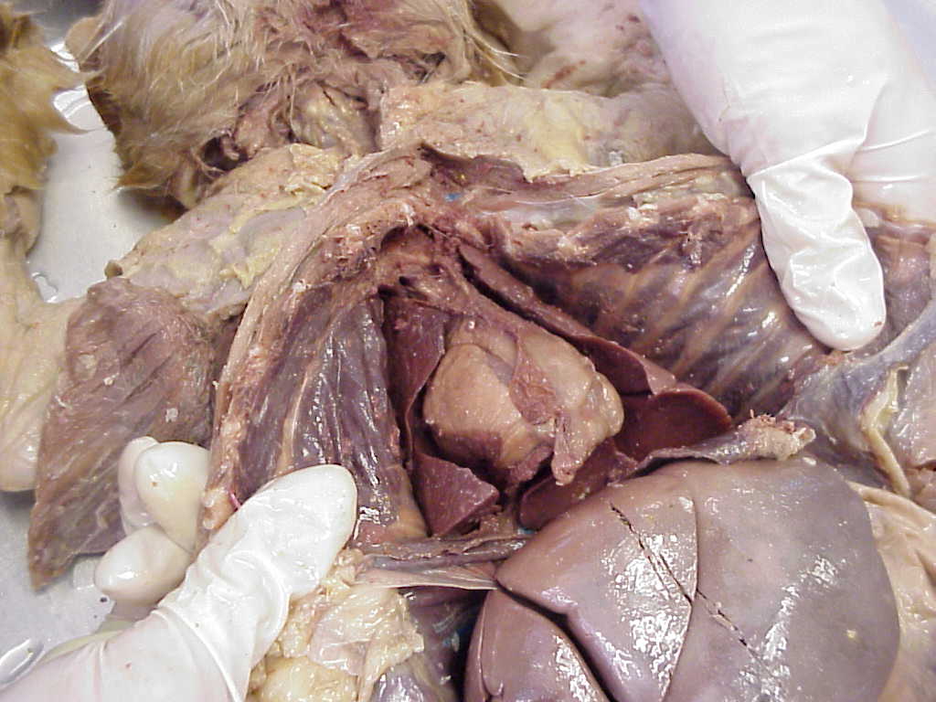
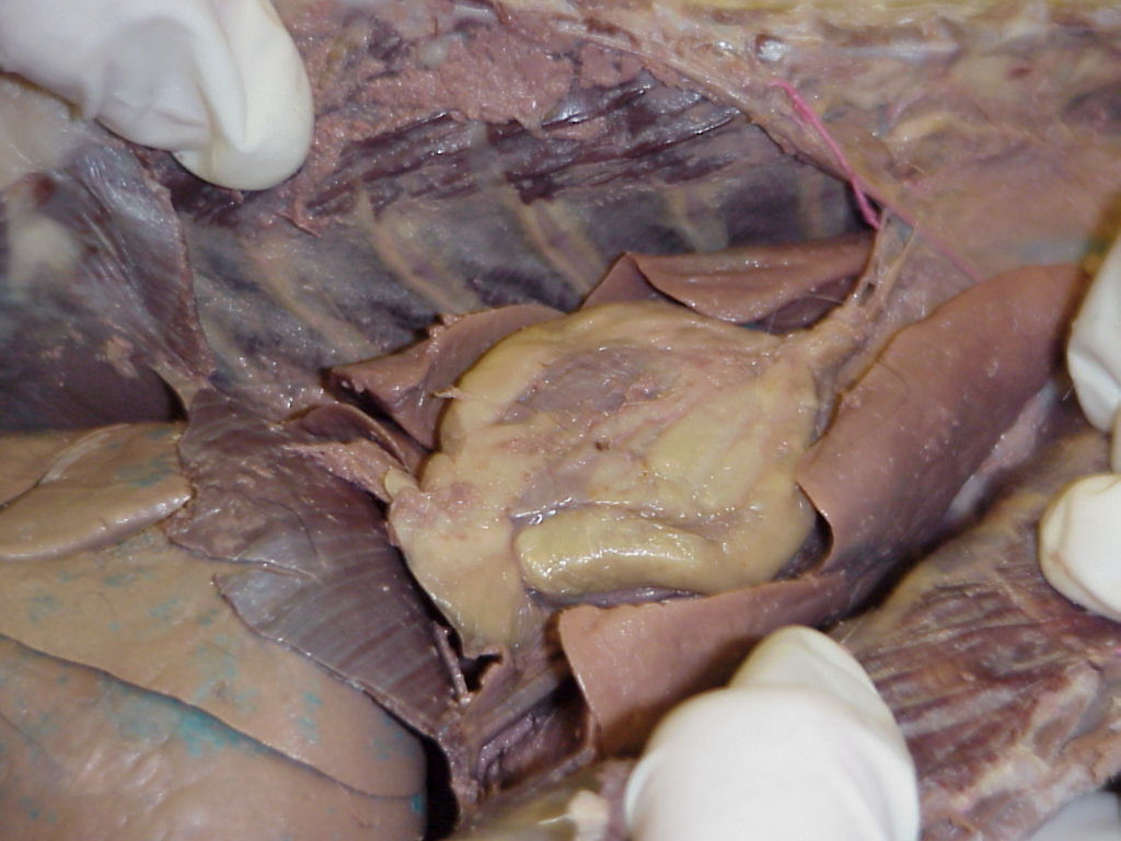
Heart, lungs and liver in exposed thoracic cavity |
Another view of the thoracic cavity |
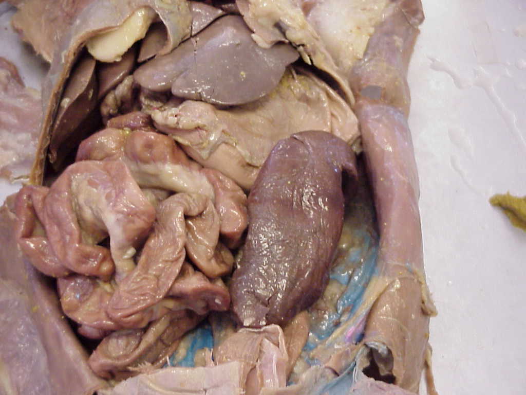
Abdominal cavity, with exposed spleen and small intestine. Stomach is superior to the spleen, and inferior to the liver. |
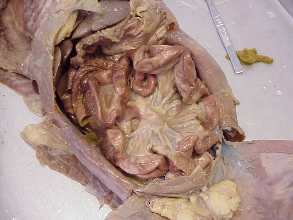
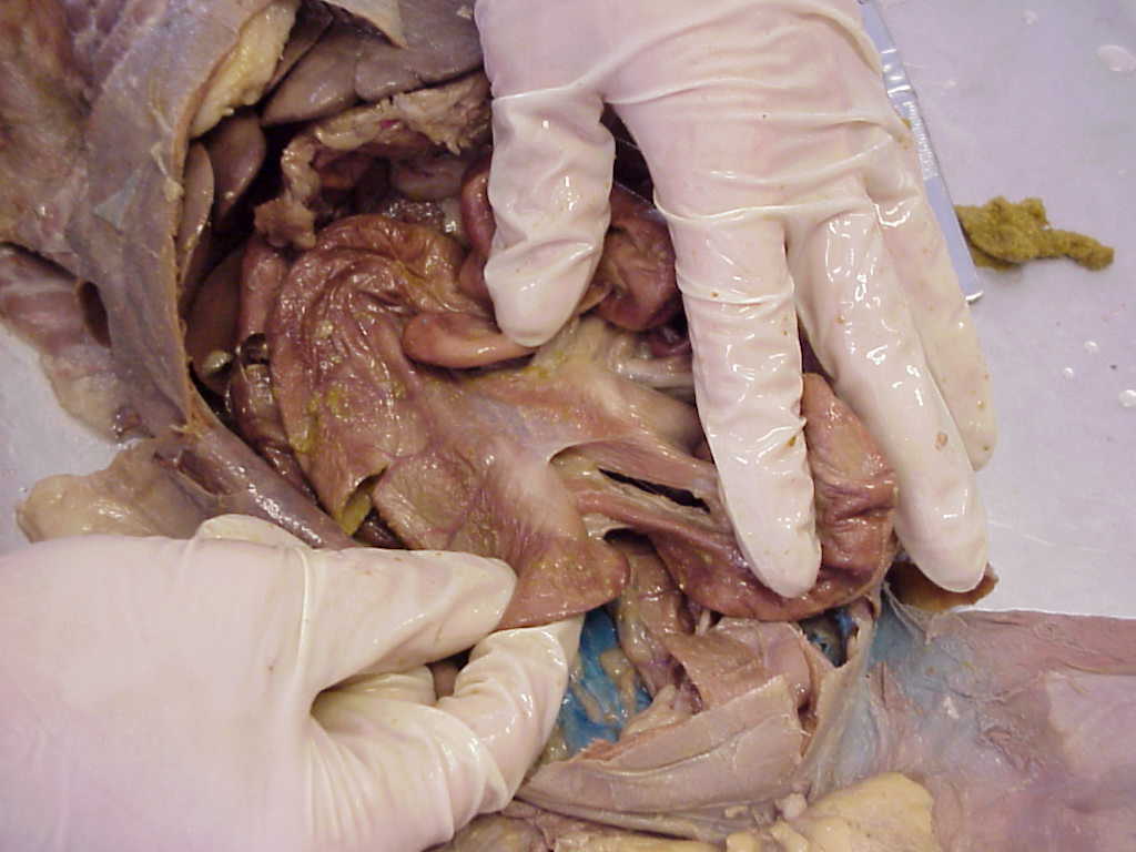
Mesentery holding together the small intestine |
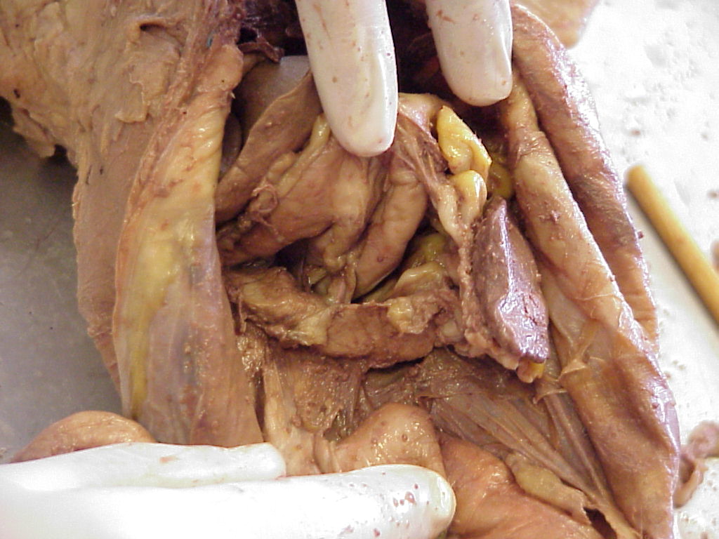
Ileocecal sphincter (valve) of the ileum and cecum |
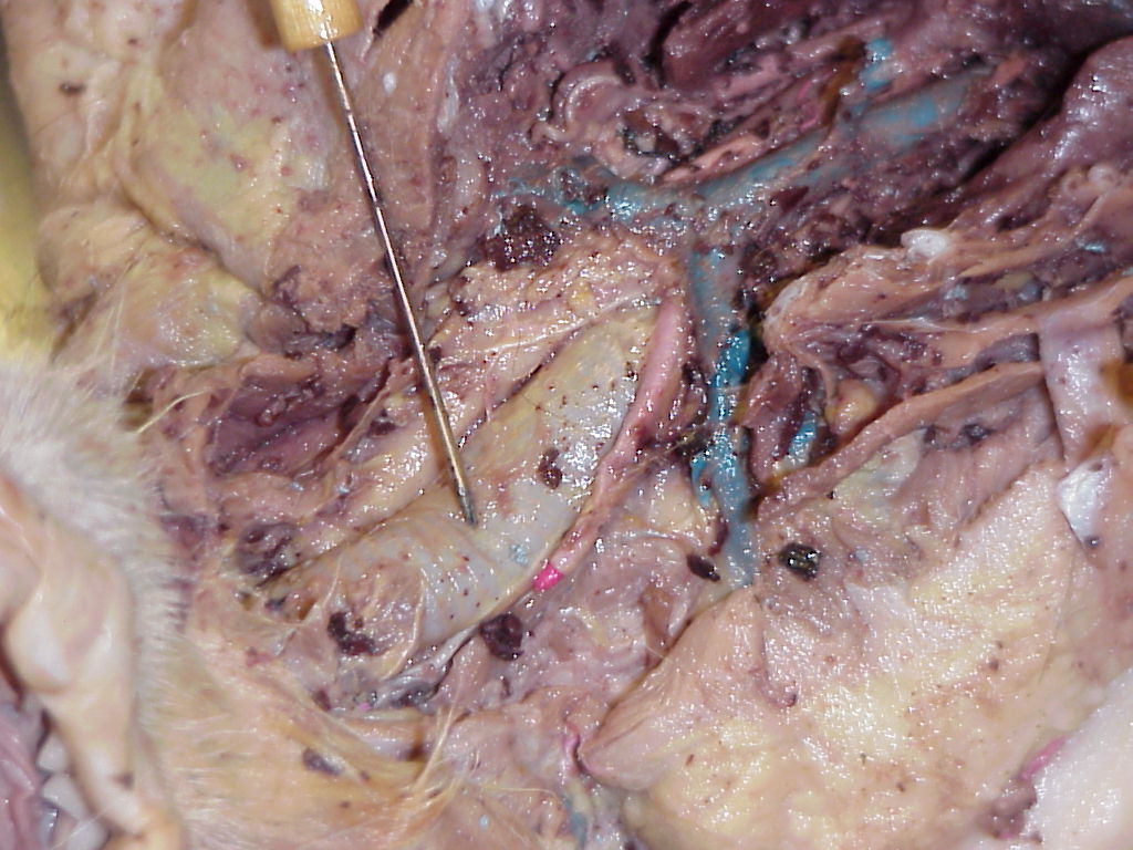
Pointer indicating trachea. Veins are superior vena cava, brachiocephalic (right brach) and right subclavian |
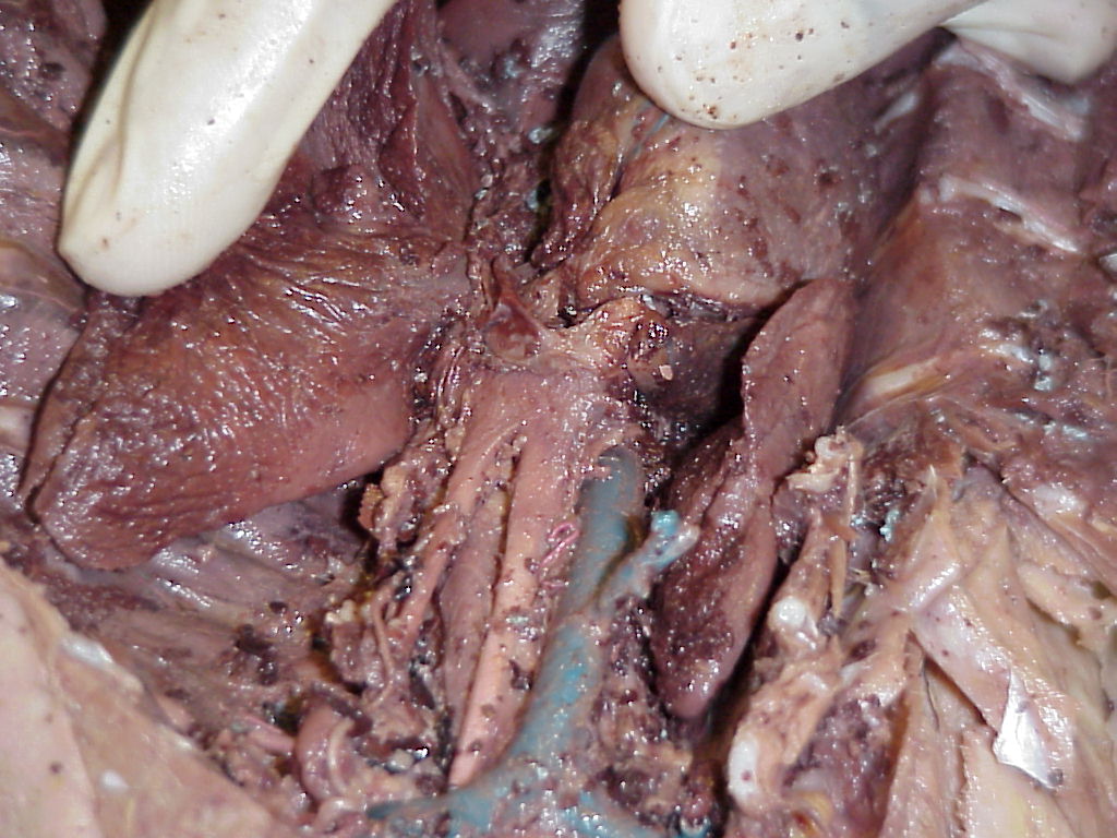
Aortic arch (from superior side). |
Aortic arch (from inferior side). |
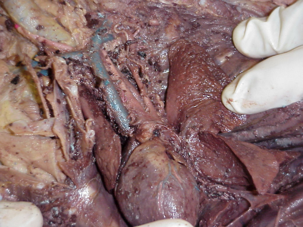
Superior vena cava (from superior side). Brachiocephalic and right subclavian veins join together to form this vein. |

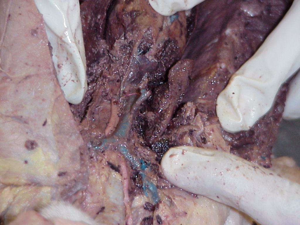
Inferior vena cava, deep to the liver. |
Thoracic cavity, showing heart with pericardium, diaphragm deep to heart and lungs. |
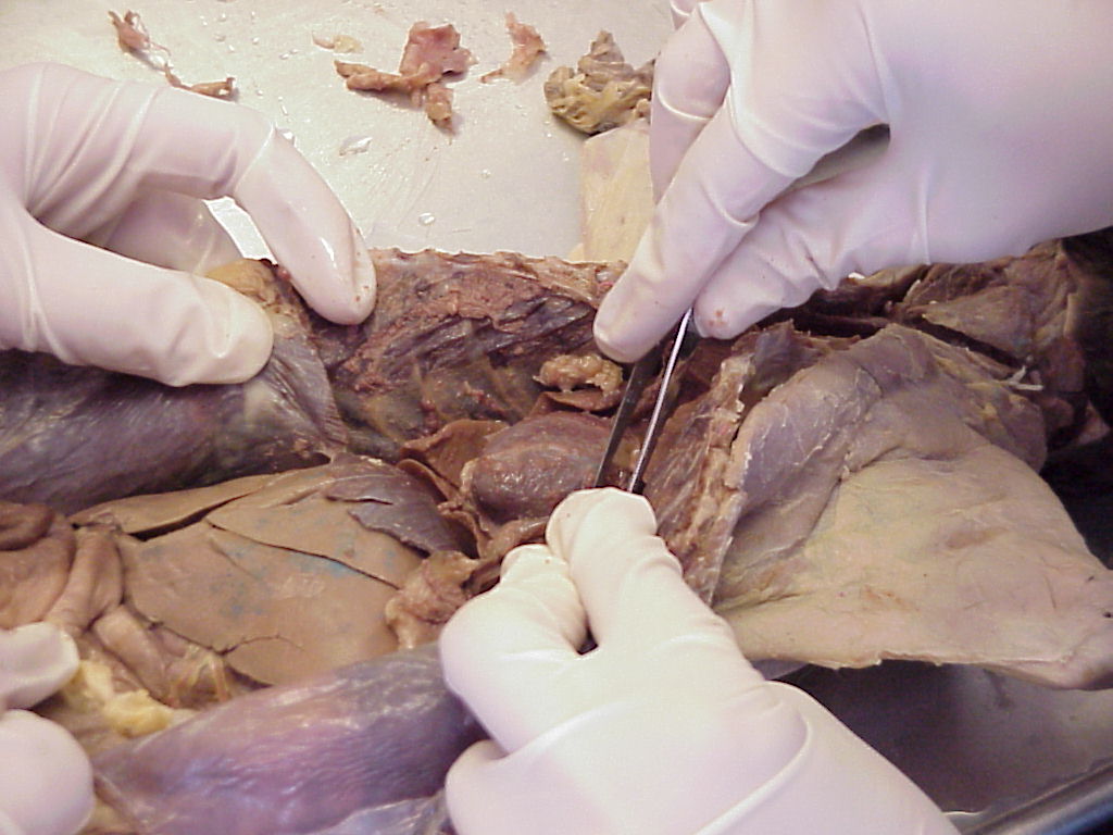
Removing the pericardium |
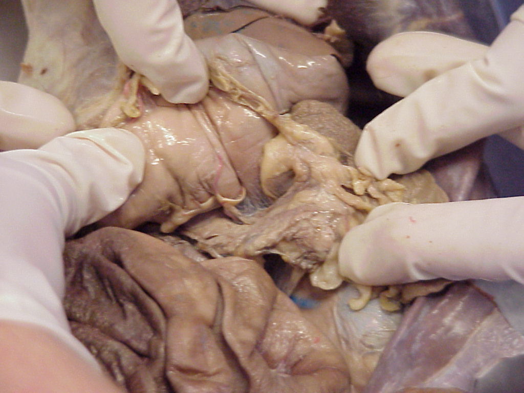
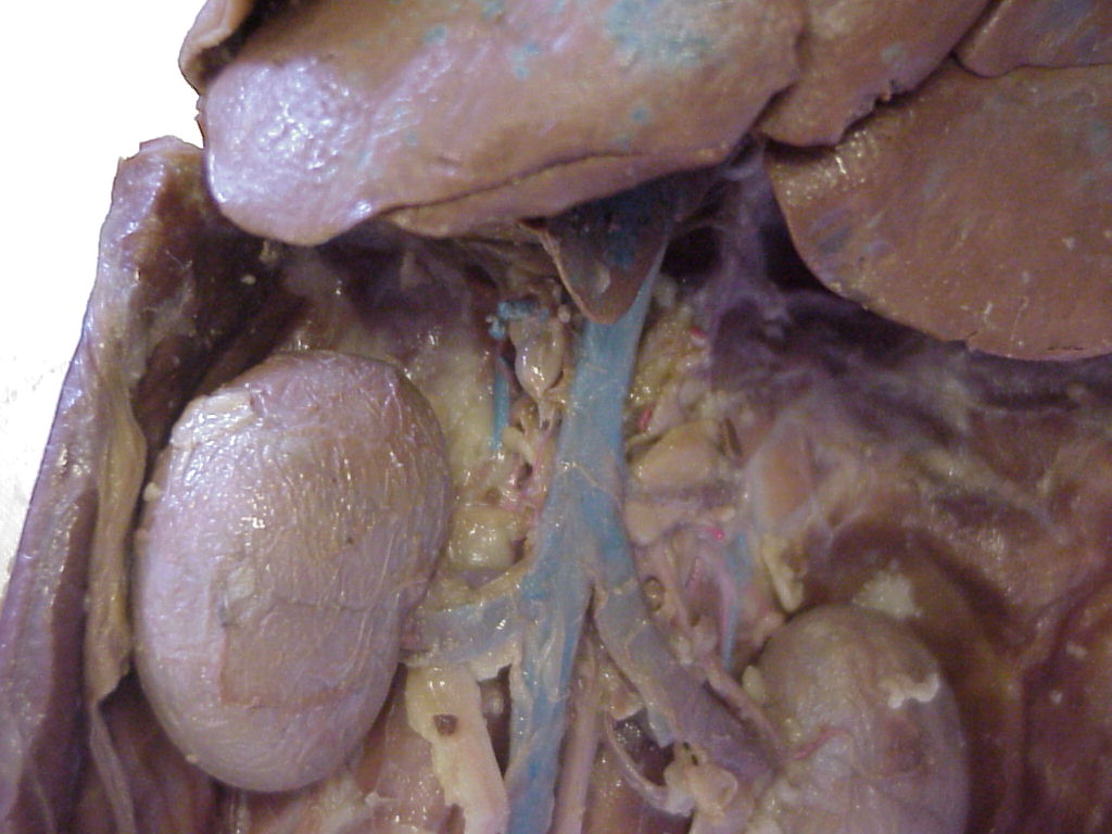
Pancreas, deep to stomach, medial to spleen |
Pancreas. |

Showing the female reproductive tract and urinary system. Note bladder, uterus, kidney, ovary, and large intestine |
Kidney, wrapped in fat pad. |
![]()
![]()
![]()
![]()
![]()
![]()
![]()
![]()