
New method for the quantification of beat-to-beat T wave temporal variability
based on inter-scale changes in wavelet transform of the ECG : Application to simulated ECG signals
.Couderc JP, Zareba W, Moss AJ
Heart Research, University of Rochester, Rochester, NY, USA
Background:
On the surface ECG, the QT segment represents the ventricular repolarization of the myocardium. Temporal variability of this segment has been shown to be a useful marker for the identification of patient with sudden cardiac death risk., The classical techniques to quantify these electrical instabilities use time (QT variability) or frequency (FFT of RT-apex variability) approaches but they both are still dependent on an accurate determination of T wave endpoints.
The primary aim of this study was to develop a new approach based on wavelet transformations (WTs), used as constant-Q filterbank, to quantify beat-to-beat temporal variability of repolarization in duration and magnitude without the need for an accurate determination of T wave endpoints. The secondary goal was to evaluate the performance of our new method under the following simulated condition imitating factors affecting ECG recordings: noisy signal and respiratory modulation.
Method:
1) Wavelet transformation: The second derivative of a Gaussian function in (1) was used as the wavelet function. The WT ![]() of the simulated ECG signal Ecg(n) relative to the basic wavelet j a(n) at scale a is defined in (2).
of the simulated ECG signal Ecg(n) relative to the basic wavelet j a(n) at scale a is defined in (2).
![]() (1)
(1) ![]() (2)
(2)
![]() and
and ![]() are the Fourier transforms of Ecg(n) and of the complex conjugate of the wavelet j a(n). TF-1 is the inverse discrete-time Fourier transform. The set of wavelets is composed of 10 logarithmically progressing scales (
are the Fourier transforms of Ecg(n) and of the complex conjugate of the wavelet j a(n). TF-1 is the inverse discrete-time Fourier transform. The set of wavelets is composed of 10 logarithmically progressing scales (![]() ) decomposing the signal following the time-frequency characteristics described in Fig. 1. The values of � varied between 0 and 3.21 with a step of 0.35. Ecg(n) and j a(n) are signals of N sample length.
) decomposing the signal following the time-frequency characteristics described in Fig. 1. The values of � varied between 0 and 3.21 with a step of 0.35. Ecg(n) and j a(n) are signals of N sample length.
For each value of a, frequency bandwidth and time duration of the analyzing wavelets are processed using (3) and (4) in which ![]() is the energy of the wavelet,
is the energy of the wavelet, ![]() and
and ![]() are the time duration and the frequency bandwidth of the wavelet, respectively.
are the time duration and the frequency bandwidth of the wavelet, respectively. ![]() and
and ![]() are the center values of the wavelet in the time and frequency domains.7
are the center values of the wavelet in the time and frequency domains.7
 (3)
(3)
 (4)
(4)
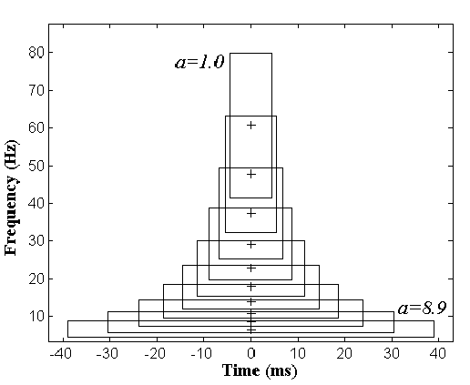
Fig. 1
: Time-frequency representation of the set of wavelets. Wavelets are centered at2) Quantification of temporal beat-to-beat variability: Our approach is based on two quantifiers of beat-to-beat temporal QT variability measuring respectively the changes in time duration and in amplitude between the scales of WT of consecutive beats. The analyzed portion of each cardiac beat must include the entire T wave and then has to be different according to the heart rate. For instance, an interval of 500 ms beginning 100 ms after the R peak can be used. Main QRS components are isolated with a Hanning window and removed from beat before processing the WT.
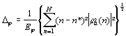
The first quantifier called Temporal Variability in Time (TVT) is based on the detection of the maximal values ma of the cross-correlation function ![]() between WTs of the beats n and n+1 for a given scale :
between WTs of the beats n and n+1 for a given scale :
{ma:![]() } (5)
} (5) ![]() (6)
(6)
The TVT value, defined in (6), is the contribution of each of the 10 scales for the shift segment estimation.
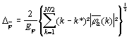
For the second quantifier called Temporal Variability in Amplitude (TVA), ![]() and
and ![]() are synchronized in the time domain using the TVT value. The WTs are then subtracted scale by scale, leading to a new time-scale representation called Mapping of Temporal Time-scale Variability (MTTV), and highlighting changes in the time-scale components between the two consecutive beats (see panels I to L in Fig. 2). The MTTV can be formulated as the following:
are synchronized in the time domain using the TVT value. The WTs are then subtracted scale by scale, leading to a new time-scale representation called Mapping of Temporal Time-scale Variability (MTTV), and highlighting changes in the time-scale components between the two consecutive beats (see panels I to L in Fig. 2). The MTTV can be formulated as the following:
![]() (7)
(7)
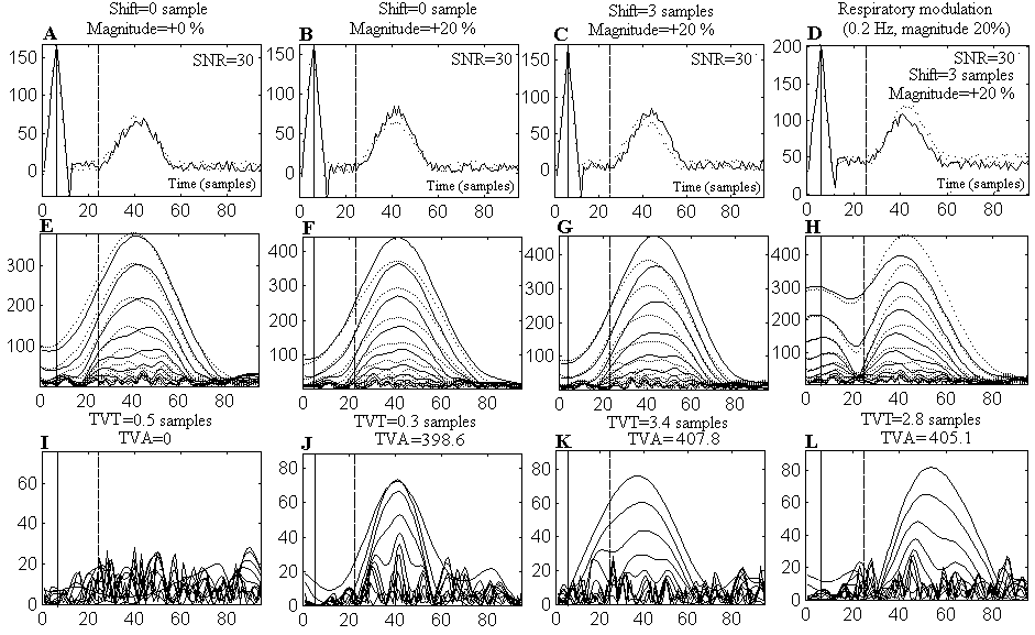
Fig. 2
: Simulated ECGs of 2 consecutive beats are superposed in panels A to D (solid and dotted curves) under different possibilities of temporal variability; their respective WTs are represented in panels E to H. MTTV are plotted in panels I to L. In the first column, no shift, no difference in magnitude, SNR is equal to 30. In the second column, difference in magnitude of 20% is simulated. In the third column, a shift of 3 samples and an increased magnitude of 20 % differentiate the 2 consecutive beats. In the last column, same temporal variability factor as in C, respiratory modulation is added. Solid verticals mark the R peak time localization. Dotted verticals indicate the onset of the analyzed segment. The TVT and TVA values are given in title of the lower panels. Panels J to L clearly show that T wave variability is highlighted in the MTTV.The computing of TVA is based on a technique derived from a previous work. This consists of tracking the evolution of �local maxima� of MTTV across scales. The technique involves four steps. (I) Detection of local maxima: all local maxima are detected on WTs scale by scale. The local maxima is defined as the point where the WT magnitude is greater than the amplitude of its nearest neighbors. (II) Removal of low amplitude maxima : an empirical threshold in amplitude is used to remove maxima due to noise. This threshold is the upper amplitude value of 90% of the maxima in all scales. (III) Localization of connected maxima : a local maximum in any of the first three scales corresponding to the lowest frequency components (4 to 14 Hz), and a local in any scale are connected if the deviation between their respective time localization is less than or equal to 5 samples. (IV) acknowledgment of connected maxima as T wave changes : T wave variability in amplitude is detected when the number of local maxima found to be connected is greater than 3.
The TVA value then corresponds to the difference in amplitude between the highest and lowest connected maxima values weighted by the number of connected maxima.
TVA and TVT are processed on each consecutive beat and the mean value is calculated on the entire lead.
3) Simulated 10-second ECGs: We simulated ECGs with heart rate of 90 bpm (256 Hz sampling frequency). Variability in magnitude and in T wave prolongation were investigated and the incidence of respiratory modulation and noise level on TVT and TVA parameters were quantified. Examples of beats from simulated ECGs are shown in panels A to D of Fig 2.
- Time duration variability was simulated using 12 levels of shift ranging from 2 to 24 by 2 sample steps.
- Usual beat-to-beat variability in the T wave amplitude is very discrete 5-20 �V for a T wave amplitude equal to 300 �V. We thus simulated T wave variability in amplitude using an increased percentage of the normal T wave. Five values ranging from 0 to 25% by a step of 5 % were used.
- The level of noise was estimated using the signal-to-noise ratio (SNR) as the magnitude of the ECG divided by the root mean square value of noise in a 100 ms window localized 80 ms after the T offset. White noise was used and the SNR values ranged from 60 to 10. ECGs with an SNR value under 25 are usually considered noisy signals.
- The frequency of respiratory modulation depends on the subject, however, it is wildly accepted that this frequency ranges between 0.16 Hz to 0.25 Hz. We thus compared TVT and TVA values for a set of ECGs with and without respiration modulation. Respiratory modulation was simulated by a sinusoid function of 0.2 Hz with a magnitude equal to 20% of the maximum ECG amplitude.
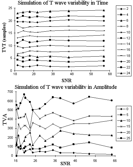
Results : Figure 3 shows the effect of different levels of noise on various degrees of repolarization variability in time (TVT; upper panel) and in amplitude (TVA; lower panel) in our simulated ECGs.
Despite seven different SNR levels superimposed on simulated repolarization variability, TVT values were equal to the magnitude of simulated shifts with a maximum standard deviation of TVA less than 1 sample (observed when noise level was high: SNR<30). These observations provide a convincing evidence that TVT is an accurate and robust quantification of T wave shifts regardless noise level.
Variability in amplitude measured by TVA values seems to be more affected by different SNR levels (SNR<25) even when there is no variability in repolarization morphology (0% line on the plot). The same phenomenon was observed for various levels of simulated T wave variability. When SNR is >30, usual ECG conditions, TVA represents accurately the magnitude of T wave amplitude variability.
Fig. 3: Description of the value of TVA and TVT parameters according to different SNR levels and following different values of simulated T wave variability in time and in amplitude.
We compared simulated ECGs with and without respiratory modulation and following the same values of temporal variability used in fig. 3. The mean values of TVT and TVA are not significantly different (p>0.1) with and without respiratory modulation, regardless the SNR level and the simulated temporal variability. The difference for the TVT values between the ECG with and without respiration modulation is less than 3 samples in any case. Respiratory modulation has no significant (p>0.1) influence on the TVA values for the set of ECG we simulated.
Conclusion: We provided evidence that WTs analysis of ECG can be used to quantify the magnitude of beat-to-beat variability of repolarization morphology, both its duration and amplitude. Our new technique quantifying inter-scale changes in the wavelet transform of ECG does not require a precise localization of T wave endpoints, what is a major limitation of existing techniques based on QT or RT-apex interval detection.
Simulation experiments showed that our new wavelet-based parameters (TVA and TVT) provide robust quantification of repolarization beat-to-beat variability in the presence of clinically relevant noise levels and respiratory modulation. This technique is under testing in clinical conditions.
Conclusion:
[1]Smith JM, Clancy EA, Valeri CR, Ruskin JN, Cohen RJ. Electrical alternans and cardiac electrical instability. Circulation 1988;77:110-121.
[2]Coumel P, Fayn J, Maison-Blanche P, Rubel P. Clinical relevance of assessing QT dynamicity in Holter recordings. J Electrocardiol. 1994;suppl 27:62-66.
[3]Merri M, Alberti M, Moss AJ. Dynamic analysis of ventricular repolarization duration from 24-hour Holter recordings. IEEE Trans Biomed Eng 1993; 40: 1219-25.
[4]Lepeschkin E, Surawicz B. The measurement of the QT interval of the electrocardiogram. Circulation 1952;6:378-388.
[5]Badilini F, Moss A J, Titlebaum L. Cubic spline baseline estimation in ambulatory ECG recordings. In: Proceedings of the IEEE/EMBS Annual conference. New-York: IEEE/EMBS, 1991:584-585.
[6]Couderc JP, Zareba W, Burattini L, Konecki J A, Moss A J. Detection of Abnormal Time-Frequency Components of the QT interval using Wavelet Transformation Technique. In: Computer in Cardiology 1997. Lund (Sweden): IEEE Computer Society Press, (in press).
[7]Morlet D, Couderc JP, Touboul P, Rubel P. Wavelet analysis of high-resolution ECGs in post-infarction patients: role of the basic wavelet and of the analyzed lead. Int J Biomed Comp, 1995;39:311-325.
[8]Badilini F. Time and frequency analysis of ST segment displacement signal in ambulatory ECG recordings. Doctoral Thesis, College of Engineering and Applied Sciences, Rochester, NY, 1991.