
Detection of Abnormal Time-Frequency Components of the QT Interval
Using a Wavelet Transformation Technique
JP Couderc, W Zareba, L Burattini, AJ Moss
University of Rochester, Rochester (NY), USA
Abstract
ECG Repolarization abnormalities are likely to be manifested by different time-frequency (TF) components but no specific technique to detect them has yet been proposed. This study aims to describe the usefulness of a time-scale technique for the analysis of the repolarization segment components. Affected patients with Long QT Syndrome (LQTS) were used as group of patients with abnormalities in repolarization.
Time-scale (TS) analysis of the repolarization interval revealed modification of the TF components with different shapes according to the leads. Comparison of ROC curves for QTc measurements and wavelet parameters demonstrates that wavelets are able to quantify T wave prolongation without the need for T endpoint determination.
1. Introduction
QT prolongation [1], T wave morphology changes [2] have been defined as non-invasive ECG parameters identifying patients at risk for sudden cardiac death. Established automatic detection of these electrical instabilities are based on time-domain measurements (QT variability) [3] or frequency-domain (FFT of RT-apex variability) approaches [4]. Unfortunately, all these techniques are still dependent on the localization of the onset and offset of the T wave, which are difficult to detect accurately. The current goal then, is to define a new technique for the quantification of the T wave abnormalities without any prior knowledge of the T wave endpoints.
The purpose of this study is 1) to describe an optimal TS technique for the analysis of repolarization components. 2) To evaluate the performance of the wavelet technique for detecting repolarization abnormalities in 12-lead ECGs of LQTS patients and Normal subjects. 3) To assess the influence of different wavelet shapes on measurements of TF repolarization components measured with the TS decomposition.
2. Methods
2.1 TF characteristics of wavelets
The six wavelets are the first five derivatives of a Gaussian function and the Morlet wavelets (Table 1) [5]:
Table 1. Basic wavelets applied to the repolarization segment.
g1 ![]() with
with ![]()
g2 ![]()
g3 ![]()
g4 ![]()
g5 ![]()
g6 (Morlet) ![]() (
(![]() =5.33 rad/s)
=5.33 rad/s)
The wavelet transformation is calculated in the frequency domain using (1) where TF-1 is the inverse Fourier transform: ![]() (1)
(1)
![]() and
and ![]() are the Fourier transform of the ECG and of the analyzing wavelets
are the Fourier transform of the ECG and of the analyzing wavelets ![]() , respectively.
, respectively. ![]() represents the set of wavelets obtained with (2) from the basic wavelet
represents the set of wavelets obtained with (2) from the basic wavelet ![]() :
: ![]() (2)
(2)
We defined, for each basic wavelet (n=1 to n=6), a set of 10 analyzing wavelets with TF characteristics ranging approximately from 5 Hz to 80 Hz (see Table 2).
Table 2. Minimal and maximal wavelet time durations and high cut-off frequencies (F max) for each set of wavelets types. The lower cut-off frequencies are equal to 5 Hz in each wavelet type. TD: time duration.
|
n |
1 |
2 |
3 |
4 |
5 |
6 |
|
Lowest TD (ms) |
43 |
36 |
48 |
40 |
50 |
67 |
|
Highest TD (ms) |
208 |
320 |
355 |
324 |
526 |
724 |
|
F max (Hz) |
64 |
78 |
80 |
93 |
84 |
81 |
|
Redundancy (%) |
72 |
55 |
49 |
43 |
32 |
0 |
In the TF representation, each wavelet can be represented by a window, the height ![]() of the window is the frequency bandwidth of the wavelet and the length
of the window is the frequency bandwidth of the wavelet and the length ![]() its time duration.
its time duration. ![]() and
and ![]() are defined by (2) and (3) and the energy of the wavelet
are defined by (2) and (3) and the energy of the wavelet ![]() by (4).
by (4).
 (2)
(2)
 (3)
(3)
![]() (4)
(4)
![]() is the Fourier transform of gn(t).
is the Fourier transform of gn(t). ![]() and
and ![]() are the center values of the wavelet in the time and frequency domains [6].
are the center values of the wavelet in the time and frequency domains [6].
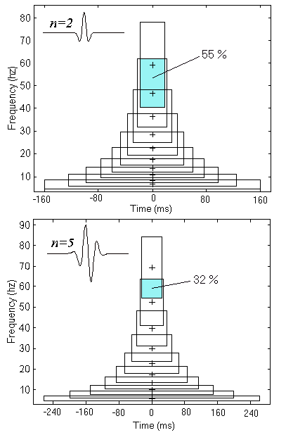
Figure 1. TF representations of the sets of wavelets from gn(t) with n=2 (upper panel) and n=5 (lower panel). Shapes of the basic wavelet functions are plotted in the upper left corner of each panel. Frequency redundancy is indicated in gray between the two first scales. Crosses mark the central frequency of the wavelet at each scale. Each wavelet is centered at t*=0 ms.
The product of ![]() by
by ![]() represents the TF area of the wavelet, which is constant for each scale. Figure 1 illustrates the time-frequency representation of the wavelet sets obtained from gn for n=2 and n=5. Wavelets show redundancy in frequency indicated by the superimposed area between each consecutive TF box. This redundancy is quantified using the percentage of common area between wavelets of two consecutive scales (see table 2 and figure 1). This common area is constant for each scale. For instance, when n=1 the common area between consecutive scales is extremely high (72%) and progressively decreases as n increases. When the Morlet wavelet is used, the set of wavelets has no redundancy in frequency.
represents the TF area of the wavelet, which is constant for each scale. Figure 1 illustrates the time-frequency representation of the wavelet sets obtained from gn for n=2 and n=5. Wavelets show redundancy in frequency indicated by the superimposed area between each consecutive TF box. This redundancy is quantified using the percentage of common area between wavelets of two consecutive scales (see table 2 and figure 1). This common area is constant for each scale. For instance, when n=1 the common area between consecutive scales is extremely high (72%) and progressively decreases as n increases. When the Morlet wavelet is used, the set of wavelets has no redundancy in frequency.
For each lead, wavelet transformations were applied to ECG segments beginning 280 ms before the R peak and ending 720 ms after this same peak, providing an entire P-QRS-T decomposition (see figure 2).
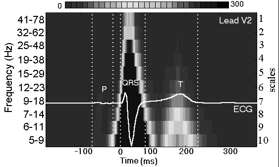
Figure 2. Example of time-scale representation obtained with g2 when applied to the median beat from lead V2 of a healthy subject. ECG is superimposed with its wavelet transformation. Vertical dotted lines indicate: onset and offset of P and QRS waves and T wave offset. Color scale was defined to highlight the components of the T wave. Scale axis is given in scale on the right and in frequency bandwidths on the left.
The wavelet transformation technique was used to evaluate TF components in the median beat of standard 10-second 12-lead ECGsin 43 Long QT Syndrome (LQTS) patients (genetically or ECG diagnosed) and 29 healthy control subjects (Normals). As expected, heart rate corrected QT duration (QTc) was significantly higher (p<.0001) in LQTS vs Normals.
Table 3. Characteristics of the populations. *: p<0.0001.
|
|
Normal (n=29) |
LQTS (n=43) |
|
Age (years) |
31 � 14 |
26 � 13 |
|
Males |
41% |
51% |
|
Heart Rate (bpm) |
66 � 15 |
62 � 10 |
|
QTc (ms) |
407 � 56 |
471 � 17* |
2.3. Mapping of time-scale abnormalities and comparison of wavelet functions
The wavelet transformation analyses are combined with statistical analysis to provide mappings of TF abnormalities (as described previously [6]). Using ANOVA method, mappings compared TF components of ECG signal between our two study populations. The mappings are 3-dimensional representations where: X-axis shows time, Y-axis shows scales or frequency bands, and Z-axis (color axis) shows p-value for comparison between groups, with N as reference. Dark colors correspond to significantly increased energy for the LQTS group, and light colors to significantly decreased energy for the LQTS group. This method allows us to precisely localize the abnormalities of the T wave simultaneously in the time and frequency domains.
We designed an algorithm automatically detecting for each mapping, sets of wavelet associated with a significant p-value (p<0.0001). The values of coefficients of all retained wavelets were summed to obtain a simple quantifier of T wave abnormalities. Then, using ROC curves, we compared the ability of each of these quantifiers to discriminate the two study populations. The efficiency of the quantifiers is based on the measurement of the ROC area. An ideal parameter would have an area equal to 100%.
3. Results
3.1 TF T-wave abnormalities
Figure 3 illustrates the results of the mapping in the 6-precordial leads, for the wavelet g2(t), uncovering different time-scale abnormalities of the T wave. The six limb leads (I to III, aVR, aVF, aVL) did not provide any additional information.
In this figure, the QT prolongation of the median beat of LQTS patients appears as a modification of T wave components in time and in frequency. Depressed and increased energy are systematically detected for each lead, with various frequency components, time duration and significance. Significantly depressed energy (p<0.0001), representing higher activity in Normal group, is centered around 270 ms after the R peak and between 5 and 48 Hz. Significantly increased energy centered around 450 ms after the R peak is also detected in all frequency bandwidths (5 to 78 Hz).
Figure 4 displays the mappings for leads V4 and V5 using wavelets for n=2, 5, and 6. These representations clearly show that the lower the value of n, the more significant the differences between the groups.
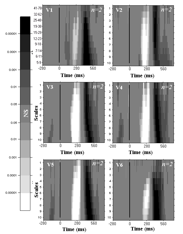
Figure 3. Time-scale mappings of the patients with LQTS onto the p-value axis with reference to the Normal subjects for all precordial leads and with wavelets from the second Gaussian derivative (n=2). The vertical lines mark the R peak. The vertical axis of the first mappings are in frequency, the others are marked using scales. Depressed and increased energies for LQTS pts are in white and in black, respectively.
Extraction of wavelet parameters reveal that the increased energy (p<0.0001) around 500ms after the R peak gave the higher ROC area regardless of the lead and the wavelet function. Figure 5 shows the ROC curves for QTc and the parameters from the six kinds of wavelet decomposition. The wavelet decomposition using low derivatives of Gaussian functions ROC curves have slightly higher values of ROC area. The Morlet wavelets have been revealed to be less efficient wavelet function (89%) for the T wave analysis because of a too long time duration. For instance, Morlet wavelet duration, at scale #10, is equal to 724 ms. This wavelet duration does not definitely separate QRS from T wave components. ROC from QTc has an area equal to 88%. The wavelet decomposition thus provides significant and robust parameters for the analysis of T wave abnormalities.
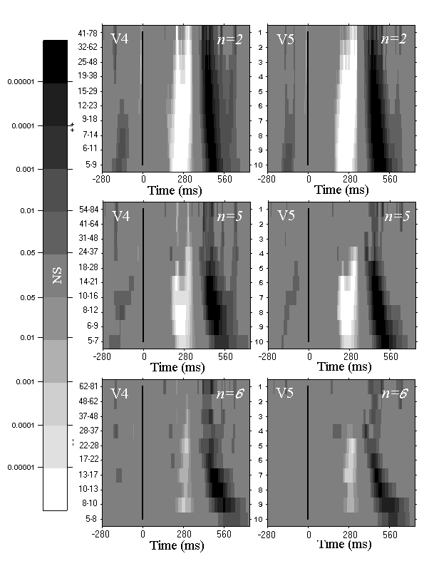
Figure 4. Mappings of lead V4 and V5 for the second and fifth Gaussian derivatives, and the Morlet wavelet (n=2, 5 and 6). Same definitions as in Figure 3.
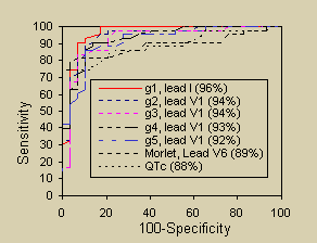
Figure 5. ROC curves for QTc and wavelet parameters. ROC areas are given in parenthesis in the legends.
4. Discussion
We demonstrated that wavelet transforms with Gaussian derivatives are able to detect repolarization abnormalities in a standard 12-lead ECG. The differences in time and redundancy in frequency contribute the most to detect abnormalities. The advantage of wavelets from the Gaussian derivative is that they have more redundancy in frequency and shorter time duration allowing enhancement of T wave abnormalities without confounding influence of QRS components. We observed that the Morlet wavelets are less effective in detecting T wave abnormalities in comparison to Gaussian wavelets. This mainly is due to the long duration of the Morlet wavelet. Wavelet technique also outperformed the standard QTc interval measurements in identifying affected LQTS patients. Our method provided insight into frequency bands affected by repolarization abnormalities. The most significant differences between LQTS patients and Normals were observed in frequencies between 5 and 48 Hz. In some leads, abnormalities between 50-78 Hz are also observed.
Wavelet transformation technique quantifying TF components of repolarization in 12-lead ECG is able to identify repolarization abnormalities without the need for determining T wave limits. Among various types of wavelets studied, the two first Gaussian derivatives provide the best identification of repolarization abnormalities. The most significant differences in frequency content of repolarization between LQTS and Normals were detected between 5 and 48 Hz.
[1] Moss AJ, Schwartz PJ. Delayed repolarization (QT and Q-U prolongation) and malignant ventricular arrhythmias. Mod Concepts Cardiovascular Dis 1982; 51: 85-90.
[2] Moss AJ, Zareba W, Benhorin J et al. ECG T-wave patterns in genetically distinct forms of the hereditary long QT syndrome. Circulation 1995;92:2929-2934.
[3] Merri M, Benhorin J, Alberti M, Locati E, Moss AJ. Electrocardiographic quantification of ventricular repolarization. Circulation 1989;80:1301-8.
[4] Merri M, Alberti M, Moss AJ. Dynamic analysis of ventricular repolarization duration from 24-hour Holter recordings. IEEE Trans Biomed Eng 1993; 40: 1219-25.
[5] Morlet D, Couderc JP, Touboul P, Rubel P. Wavelet analysis of high-resolution ECGs in post-infarction patients: role of the basic wavelet and of the analyzed lead. Int J Biomed Comp, 1995;39:311-325.
[6] Couderc JP, Fareh S, Chevalier Ph, Fayn J, Kirkorian G, Rubel P, Touboul P. Stratification of time-frequency abnormalities in the signal-averaged high-resolution ECG in post-infarction patients and congenital Long QT Syndrome. In: J. Electrocardiol.1996, 29 (suppl):180-88.
Address for correspondence:
JP Couderc, Heart Research, University of Rochester,
601 Elmwood Av. Box 653, Rochester, NY, 14620 USA
E-mail: [email protected]