
Contribution of the Wavelet Analysis to the Non-Invasive Electrocardiology
Jean-Philippe Couderc, PhD, and Wojciech Zareba, MD, PhD
Heart Research Follow-up Program, University of Rochester, Rochester, New York, USA
Running Title: Wavelets in non-invasive electrocardiology
Address for correspondence:
Jean-Philippe Couderc
Heart Research
601 Elmwood Avenue, Box 653
Rochester, New-York 14642
USA
Tel: (716) 275-1096
Fax: (716) 473-2751
E-mail: [email protected]
Today, various digital-signal-processing methods are applied to the ECG to identify, extract and analyze the different ECG signal components. In this large set of signal-processing tools, a new technique called wavelet transformation appears to be a promising method describing time and frequency characteristics of ECG waves. This article aims to provide an overview of the wavelet technique applied to the area of quantitative electrocardiology without describing mathematical details of the wavelet theories, that can be found elsewhere1-3.
The first part of the article will give some rationale for the utilization of new ECG processing tools and a conceptual definition of the wavelet transformation. The second part will describe the contribution of the wavelet transformation in quantitative electrocardiology. This technique will be discussed and compared to the classical techniques using time-domain and frequency-domain measurements.
Rationale for developing new ECG-processing technique
In quantitative electrocardiology, the classical approach is to use a time-domain method, which has multiple application (standard ECG measurement, heart rate variability, and dispersion of repolarization) 4,5. However, measuring amplitude and duration of ECG waves using time-domain methods is not always sufficient to describe all features of ECG signal. For example, identification of late-potentials located inside the QRS complex can not be accomplished with the time-domain methods. Similarly, time-domain analysis of heart rate variability provides insight into overall RR variance and parasympathic influence only. Sympathetic regulation can not be evaluated based on time-domain measures of heart rate variability6. In such circumstances techniques using frequency-domain and time-frequency domain were found to be useful7-10.
The frequency representation of a signal can be obtained using different techniques including the Fourier transformation, and the autoregressive method. The most frequently used in electrocardiology is the Fast-Fourier Transform (FFT) that is able to decompose any temporal signal (theoretically this signal should be deterministic and periodic) in an infinite set of sinusoid functions. This set of sinusoid functions is then represented in the frequency space using the amplitude and the phase of each of these functions. The FFT thus provides a link between the time representation of a signal (in seconds) and the frequency representation (in Hertz or cycle/second).
As the digitized ECG is a finite signal, its boundaries are usually abrupt. These abrupt cuts of the signal make it discontinuous. This introduces a smearing (decrease and spread) of all the estimated frequency peaks. In order to avoid this, the calculation of the FFT is applied to the windowed ECG. The windowing aims to smoothly decrease the boundary of the ECG signal to zero, removing its discontinuity. The limitation of this approach is that windowing reduces the frequency resolution and therefore lowers the quality of the estimation of the ECG signal frequencies.
Another unavoidable limitation of the Fourier transformation for the ECG analysis is that this technique does not provide insight into exact location of frequency components in time. The frequency content of the ECG varies in time; the QRS complex is a high frequency wave whereas the T wave contains low-frequency components. Therefore, the need for an accurate description of the ECG frequency contents according to their location in time is essential. Utilization of time-frequency representation in quantitative electrocardiology is thus justified. This kind of representation provides insight into three dimensions of the ECG signal: the time, the frequency and the amplitude (figure 1).
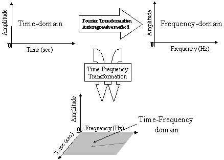
Figure 1: Time-frequency domain represents a combination of time-domain and frequency-domain characteristics of the ECG signal.
The widely used technique is the short-term Fourier transform, in which a set of FFT is calculated in a successive and overlapping windowed portion of the signal. The location and the length of this portion of the ECG define the time precision and the location in time for the spectral estimation. Several methods for late potential detection are based on this technique such as the spectro-temporal mappings and the spectral turbulence analysis 11,12. However, the short term Fourier transform has an important drawback. Its time-frequency precision is not optimal and definitely not adapted to the late-potential detection. This time precision could be increased in shortening the window but this would decrease the frequency resolution. Some other time-frequency transformations as the Wigner distribution have better time-frequency resolution than the short term Fourier transform. Among these various time-frequency transformations, the wavelet transformations also called time-scale transformations, is gaining a particular interest in quantitative electrocardiology.
The wavelet transformations and its applications
Conceptual definition of the wavelet transformation
The wavelet transformation is based on a set of analyzing wavelets allowing the decomposition of the ECG signal in a set of coefficients. Each analyzing wavelet has its own time duration, time location and frequency band. The wavelet coefficient resulting from the wavelet transformation corresponds to a measurement of the ECG components in this time segment and frequency band. The so-called wavelet coefficient is obtained following two steps: the multiplication of the analyzing wavelet times the ECG signal, and the measure of the area under the resulting curve (see figure 2). The wavelet transformation can be implemented in two techniques: redundant or non-redundant. In the redundant wavelet transformation, the set of analyzing wavelets includes wavelets with superimposed time location and/or common frequency bands. An example of redundant wavelet transformation of the ECG from healthy subjects is illustrated in the left panel of the figure 3. In the non-redundant transformation, the set of analyzing wavelets contains wavelets without superimposed time location and common frequency band.
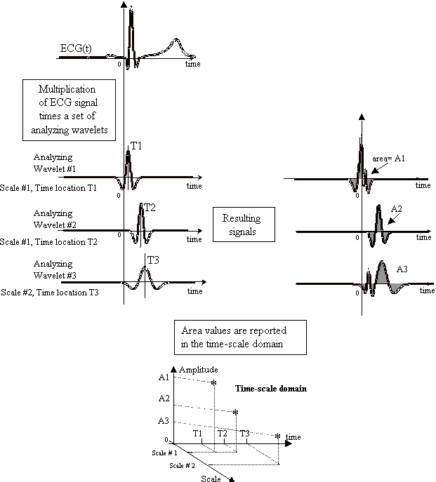
Figure 2: The wavelet-coefficient calculation is illustrated using a set of analyzing wavelet from the �Mexican hat� wavelet and the ECG signal from a healthy subject. The analyzing wavelets are first multiplied by the ECG signal. Then the wavelet coefficients are calculated using the area under the resulting curves. The area values are then plotted in the time-scale domain providing the three-dimensional representation of the signal.
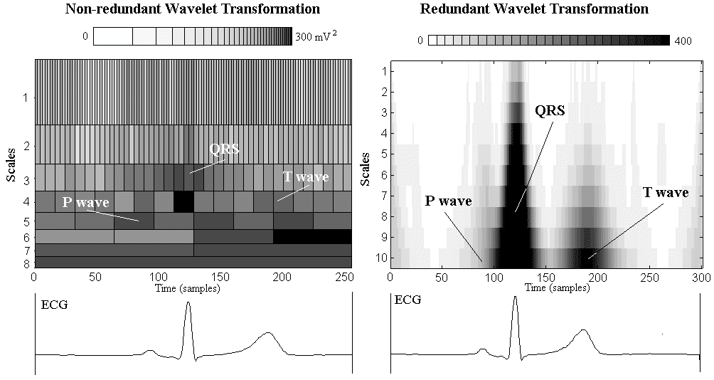
Figure 3: Examples of wavelet transformation of the median ECG from lead V6 of a healthy subject. In the left panel, a non-redundant wavelet transformation has been used (Meyer's wavelet following 8 scales). The right panel illustrates a redundant transformation obtained with a set of 10-scales 'Mexican Hat' wavelet. Scales describe frequency components of ECG with high values reflecting low frequencies and low values high frequencies. For both wavelet decomposition the amplitude of the wavelet coefficients are represented using a color scale. Darker the color, higher the coefficient value. When the non-redundant wavelet is used the wavelet coefficient is in mV2. Whereas the redundant-wavelet coefficients are dimensionless.
Redundant and non-redundant wavelet transformation have different objectives. Redundant transformation can be used to highlight ECG components and is able to accurately localize these components in the time and the frequency domains. This transformation requires a large amount of calculus and of data. On the other hand, the non-redundant transformation is faster to process because it uses a low number of coefficients (sometimes it is used for the data compression) but it does not provide accurate time location and frequency identification of the ECG components.
The set of analyzing wavelets is designed from a basic wavelet called the �mother wavelet'. This set is obtained by dilating or contracting the mother wavelet using a scale parameter that describes the wavelet dimension in both time and frequency domains. The mother wavelets are short oscillating waves designed using mathematical functions satisfying several conditions including a mean value equal to zero and boundaries quickly converging to zero. There are many kinds of wavelet functions including Morlet wavelet, Gaussian derivatives, Daubechies, Mallat, Meyer wavelets that can be applied to ECG signal processing (some of them are represented in the figure 4).
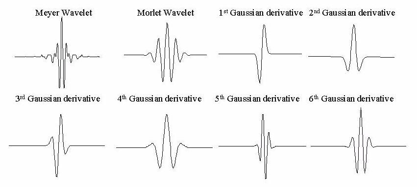
Figure 4:Frequently used wavelet functions in ECG signal processing.
The wavelet is located in time using a time parameter. Therefore this technique is also called time-scale transformation. The dilatation or contraction of the mother wavelet has influence on the time and frequency characteristics (see figure 5). When the wavelet is dilated (longer in time), its frequency bandwidth is narrowed and centered in low frequencies. On the other hand, when the wavelet is contracted (shorter time duration), its frequency bandwidth is widened and centered in higher frequencies. Thus, when the wavelet analyzes slow waves as the T wave, longer wavelets are needed and frequency resolution is good. Whereas with rapid waves, like the QRS complex, shorter wavelets provide better signal time description: time-resolution is good but frequency resolution is poor. As a microscope can focus on specific details of a slide, the wavelet shape can be adapted to focus the analysis on specific components of the ECG.
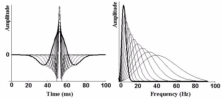
Figure 5: An example of a set of analyzing wavelets from the 2nd Gaussian derivative ('Mexican Hat') is plotted. The wavelets are represented in both the time (left panel) and the frequency (right panel) domain. The mother wavelet is drawn with a bold line in the time and frequency domains. Then for each of the ten values of the scale parameter the wavelets are plotted with same time location. The corresponding frequency representation of this set of wavelet is given. The longer the wavelet duration, the shorter its frequency bandwidth centered in the lower frequencies.
There are no established rules as to the choice of wavelet functions. A cautious and still exploratory approach is to test different wavelets and then to compare their efficiency in highlighting specific ECG information 13. In the following section we will describe several examples of wavelet applications in electrocardiology. The contributions of wavelets to non-invasive electrocardiology in comparison with classical approaches will be addressed.
Application of the wavelet transform in electrocardiology
ECG compression
Increasing use of computerized ECG processing systems requires effective ECG data compression techniques which aim to enlarge storage capacity and improve methods of ECG data transmission over phone and internet lines. The wavelet compression methods described in 1992 provide a robust technique suited for detecting and removing redundancy in the signal 14. The few publications available on this topic suggest that the ECG data compression using wavelets could decompose the ECG without any redundancy and provide high compression ratio and high quality reconstruction of ECG signal 14-17. According to these preliminary reports, wavelet-based compression seems to be more efficient than the classical compression methods.
ECG pattern recognition
One of the crucial steps in the ECG analysis is to accurately detect the different waves forming the entire cardiac cycle. Some studies 18-20 aim to design effective methods for detection and classification of ECG waves. For instance, Li et al.19 developed a wavelet-based classification method that correctly identifies 99.8% of ECG waveforms from the MIT/BIH arrhythmia database. Some authors have also shown that the wavelets, configured following a wavelet network, provide efficient extraction for discriminating between normal and abnormal cardiac pattern 21,22. Even if many algorithms have already been defined for ECG pattern recognition, the wavelet transformation seems to offer a new approach worth investigating, especially in areas of limited performance of current techniques, like P and T-wave recognition.
ST segment analysis in ischemic patient
The wavelet technique has also been used for the evaluation and monitoring of ischemic ECG changes 23,24. Mac Leod et al.24 used wavelet technique for identification of the ECG changes resulting from acute coronary artery occlusion and reperfusion observed during PTCA procedure 24. This study demonstrates that the wavelets are able to identify specific detailed time-frequency components of ECG signal, which are sensitive to transient ischemia and eventual restoration of electrophysiological function of the myocardial tissue. Since ST segment monitoring is frequently used in standard exercise testing, and Holter ECG recordings, the wavelet technique might enhance diagnostic performance of those routine ECG methods.
Heart Rate Variability
The FFT method, frequently used to evaluate heart rate variability (HRV), has inevitable limitations related to spectral leakage caused by abrupt changes at the boundary of the HRV signal. In addition, FFT introduces inconsistent spectral components of the tachogram. To overcome this problem, the tachogram can be windowed. The windowing induces modification of statistical properties of the signal called stationarity that is required to find good correspondence of energy between frequency and time domains. With the windowing, the stationarity of the signal is lost and the reliability of the spectral estimate is decreased.
In contrast to the FFT method, the wavelet HRV approach is free of any assumption about the stationary of ECG signal and permits the localization of changes in the characteristics of HRV without loosing time and frequency information 25,26. The wavelet transformation approach should also be preferable to the non-parametric technique because of no requirement about the order of the model. Wiklund et al.25 suggest that adapted wavelet transform methods can be used to detect transient changes and hence characterize and quantify both tonic and reflex autonomic activity. Wavelets are an interesting and promising alternative for the FFT or autoregressive analysis of the HRV.
High�resolution signal-averaged electrocardiography (HRECG)
Because the efficiency of the time-domain late-potential detection is limited to the terminal portion of the QRS complex and is also affected by inaccuracies of QRS-end detection, frequency-domain methods have been investigated 7,27-30. Cain and coworkers published several studies 7,27 using the FFT of the signal-averaged high-resolution ECG for identifying high-frequency components at the end of the QRS complex. The amplitudes of the peaks of the spectrogram between 20 and 50 Hz were significantly increased for post-infarction patients prone to ventricular tachycardia. In the later studies, Kelen et al.and others28-30 showed that this method was very dependent upon the interval location and interval duration for the spectrogram calculation. Analyzing time-frequency representation was then proposed and tested by several authors with FFT-based time-frequency representations 11,12,31. Abnormal high-frequency components were identified in the post-infraction patients prone to ventricular tachycardia. However, the FFT-based time-frequency representation did not provide clinically relevant improvement for the late potential detection in comparison with the time-domain approach. Indeed, high-frequency components of the late potentials were buried in the estimated frequency components of the entire QRS complex.
The HRECG analysis is the field of research most actively seeking to benefit from the wavelet signal-processing technique. In 1989, Meste et al.32 applied for the first time, the wavelet transform to 5 KHz sampled ECGs. Subsequently, they used the Meyer wavelet for the detection of the late potentials 33. The first quantitative analysis of the HRECG using wavelet transformation was described by Dickhaus et al.34, who identified significant differences in HRECG between post-infarction patients with ventricular tachycardia and healthy subjects.
A different approach was used by Shinnar and Simson 35 who examined the local scaling behavior of the ECG wavelet transformation. Patients without ventricular tachycardia produced ECG wavelet transformation with relatively constant slope, while patients prone to ventricular tachycardia produced ECG wavelet transformation with multifractal behavior. Morlet et al.36 designed a wavelet-based method for the detection of irregular structure, or singularities in the HRECG, consisting of an algorithm for tracking the evolution of the so-called local maxima of the wavelet transform across scales. This method is based on the detection, the connection and the acknowledgment of the connected maxima as signal singularities. It provides an increase in sensitivity and specificity for detecting late potentials in comparison with the results from the time-domain approach. Couderc et al.37,38 and Rubel et al.39 reported studies using non-redundant wavelet-decomposition of the HRECG for the accurate description of the time-frequency components of late potentials without the need of QRS-endpoint localization. In populations of post-myocardial infarction patients with and without sustained ventricular tachycardia, new quantifiers quantifying the energy of the high-frequency components (125-250Hz) from the QRS-ST complex were defined. These new parameters had higher discriminative power than the time-domain parameters. More recently, Reinhardt et al.40 and Sierra et al.41 from the same group published two wavelet-based approaches for HRECG analysis. Reinhardt 40 studied the value of a wavelet correlation function providing a type of spectral turbulence or scale turbulence quantification. The authors reported more spectral changes in the QRS complex of anterior myocardial infarction than in inferior myocardial infarction. Moreover the combination of time-domain analysis of late potentials and wavelet correlation functions increased the prognostic value of the ECG for predicting cardiac events after MI.
Sierra et al41, on the other hand, used the total energy in a seven-frequency band of an orthogonal wavelet decomposition of the HRECG and applied it to a prospective study population that included patients with a history of arrhythmic events. The combination of relative energy and time-domain parameters enhanced the positive predictive values when compared with applying each method alone. The author provides good illustration of the benefit of the orthogonal WT of the HRECG for the detection of abnormal time-scale patterns characterizing post-MI patients with arrhythmogenic risk.
However, no specific wavelet techniques have yet demonstrated a clinically relevant advantage (of wavelet) over the classical Simson's method for late potential detection. The major contribution of the wavelet to late potential detection is that the wavelets also seem to be able to detect abnormal intra-QRS potentials 42. An example of wavelet transformation of HRECG is presented in the figure 6. This approach is of particular value in the detection of late potentials in patients with anterior infarction and those with bundle branch blocks.
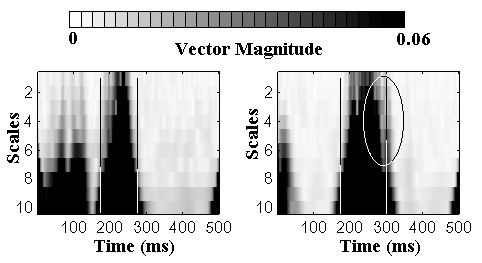
Figure 6: Examples of redundant wavelet transformations of the vector magnitude from two post-infarction patients without (left panel) and with (right panel) sustained ventricular tachycardia. The two vertical lines mark the onset and the offset of the unfiltered QRS complex. The ellipse emphases the time-frequency area with abnormal high-frequency components between 89 and 230 Hz and between 80 and 130 ms after the onset of the QRS complex.
Analysis of ventricular repolarization
Previous reports describing benefits of wavelets in ST segment monitoring (PTCA) indicate that other components of the repolarization (ST-TU complex) could be evaluated with this new signal processing technique. Our preliminary observations of the long QT syndrome gene carriers and non-carriers demonstrate that these two groups differ not only in the repolarization duration but also in specific time-frequency characteristics 43. The practical benefit of the wavelet-based ECG approach is that T wave abnormalities can be assessed without the need for T wave endpoint identification 13,43. Describing time-frequency components of the repolarization signal might provide a new approach for diagnosing patients with repolarization abnormalities. The wavelet ECG quantifiers could thus be a new alternative to time-domain analysis of the repolarization segment, especially for diagnosing the long QT syndrome in patients with borderline QT duration and for quantifying heterogeneity of repolarization44.
Summary
Time-domain analysis of the ECG is the simplest approach for detecting and quantifying the different ECG waves. However, efficacy of this approach is often limited by the inaccurate definition of ECG wave endpoints (end of the QRS, T wave location). More complex signal-processing tools, i.e. simultaneous time and frequency domain techniques, were developed to provide more accurate representation and identification of the ECG potentials. The wavelet transformation is a new promising technique in non-invasive electrocardiology providing improved methods for late-potential detection, HRV analysis and evaluation of the repolarization segment abnormalities. The benefit of the wavelet transformation lies in its capacity to highlight details of the ECG signal with optimal time-frequency resolution. Since the application of wavelet transformation in electrocardiology is relatively new fields of research, many methodological aspects (choice of the mother wavelet, values of the scale parameters) of the wavelet technique will require further investigations in order to improve the clinical usefulness of this novel signal processing technique. Simultaneously diagnostic and prognostic significance of wavelet techniques in various fields of electrocardiology needs to be established in large clinical studies.
References